Vitality, Medicine & Engineering Journal

AND option

OR option

UNIVERSAL DESIGN AS A PARADIGM FOR PROVIDING HEALTH INTERVENTIONS FOR OLDER ADULTS 2: CASE STUDY: BRANDY TAYLOR’S MUG


Peter J. Snyder


Corresponding to: Peter J. Snyder, Ph.D. Department of Art & Art History, College of Arts & Sciences, and Department of Biomedical & Pharmaceutical Sciences, College of Pharmacy, The University of Rhode Island, Kingston, RI, USA, pjsnyder@uri.edu

Care Weekly 2021;5:17-19
Published online June 2, 2021, http://dx.doi.org/10.14283/cw.2021.4


Hand strength and dexterity begin to diminish as we age, and holding everyday items like a traditional coffee mug can become difficult. The inability to properly grasp a mug of hot liquid is a common cause of burn injuries for the elderly. Brandy mitigated this home safety risk by designing a new mug that, although comfortable for elderly hands to hold, would be a pleasure for anyone to enjoy.
Brandy chose to design for the older range of the baby boomers (those born around 1946) – or people who were in their twenties during the 1960s, and so she created an inspiration board (Panel 2-2) that depicted the lifestyle of the user group she chose to study (young adults in the 1960s). To help us understand the context for her design, her image board (Panel 2-2) examined the home and family life of those she imagined using her mugs. She created a color palette, choice of materials, and a visual feeling that would be especially attractive to older adults who were raised in this era. The earth-toned colors, natural materials, and textures that she found – all dating from popular design elements in the 1960s – clearly shone through in her finished product.

Panel 2-2

Concurrently with her consideration of aesthetic choices, Brandy looked at existing hot and cold liquid containers developed specifically for the elderly, mugs from the 1960s, 70s, and 80s. She drafted several revisions of her product statement, resulting in this final description of her initial concept (Panel 2-3):
“to create a pair of mugs that are easier for people with decreased hand strength to use that are also attractive and modern looking.

Panel 2-3

To amplify on the initial concept that Brandy listed (Panel 2-3), she expanded by stating that “since the handle seems to be the most problematic aspect of mugs, why does a coffee cup even need a handle?” Creating a mug with a ceramic interior that fits into a wooden base would not only eliminate the need for a handle, but it would keep liquid warmer for a longer period of time. Additionally, a ceramic interior would allow for easy clean up. Many cups currently on the market for people with arthritis are made of plastic and need to be washed by hand, which means lots of frustration and they often have either a ‘clinical’ appearance or they may look like “sippy cups” for small children. And, her goal was to “complete a well-rounded product that solves these problems, by relying on user-testing, protoypes and surveys to iterate and refine [her] ideas.”
It was Brandy’s careful attention to understanding the user experience, to choosing form and shape based on simple aesthetics, but to refine those shapes to meet the need of the target user group, that we appreciated so much in her work.


Identification of Basic Forms for Consideration

Something as simple as a drinking mug. How hard could this possibly be? There are actually a wealth of choices and decisions to make. Brandy first started with initial concept sketches (Panel 2-4), and a subset of these were then ‘standardized’ in order to compare across shapes in a more uniform manner (Panel 2-5).

Panel 2-4

Panel 2-5

Her forms were then further refined based on an assignment to use of the “Golden Ratio” theorem, and its attendant “golden pentagram,”. This is a mathematical statement of perspective that may date back to the Ancient Greek mathematician, Euclid, and that has been relied on by countless architects, builders, designers and even musicians for the past 2,400 years (Panel 2-6).

Panel 2-6

Her work on this step resulted in 10 final forms that she chose to compare in an opinion survey (Panel 2-7).

Panel 2-7

With her 10 candidate forms selected, she collected opinions via survey of her classmates, her professors, and at least 20 older adults whom she became acquainted with as a result of repeated class excursions to a local assisted care living facility. This allowed her to arrive at two final forms that were perceived to be BOTH aesthetically pleasing as well as ergonomic (easy to grasp, hold and use without accidental spills) (Panel 2-8).

Panel 2-8


Customizing her Chosen Form(s) For her Target User Group

Brandy took her two candidate designs, and further refined them based on her study of the human hand (1), and the hands of the older adults she came to know as a result of repeated class visits to a local assisted care living facility. Her goal was to create a final form that was easy to grip, to hold, and to use without relying on fully intact upper motor strength and dexterity. She created many such forms in Styrofoam and in Plaster-of-Paris, and she brought them to the assisted care facility for field-testing (Panel 2-9).

Panel 2-9

Her final form, brought forward for initial prototyping towards the end of her 14 week class term, included a 3D-printed ceramic insert that was watertight, had excellent insulation properties, was resistant to accidental breakage, and dishwasher safe. This insert was paired with a wood shell that allowed for eliminating the typical mug handle altogether, dispenses heat evenly, and also serves as a good insulator (Panel 2-10).

Panel 2-10

Brandy’s design and testing process clearly met her stated goal of designing a hot liquid mug that anyone would enjoy using, that is visually pleasing, and that allows for safe consumption of hot liquids by older adults with restricted grip strength or with arthritic conditions, but at the same time does not suggest any connection to medical or nursing home care. Imagine a home with objects as thoughtfully designed as this simple mug? This is a product that both encourages safety and independence, but is also one that avoids or removes any hint that the target user group requires special attention because they are damaged or impaired.


Conflicts of Interest
None


Reference

1. Dreyfuss, H. (A.R. Tilley, Ed.) (2002). The Measure of Man and Woman: Human Factors in Design, Vol. 1. New York, NY: Wiley. (Panel 2-9, left)

UNIVERSAL DESIGN AS A PARADIGM FOR PROVIDING HEALTH INTERVENTIONS FOR OLDER ADULTS 1


Peter J. Snyder


Corresponding to: Peter J. Snyder, Ph.D. Department of Art & Art History, College of Arts & Sciences, and Department of Biomedical & Pharmaceutical Sciences, College of Pharmacy, The University of Rhode Island, Kingston, RI, USA, pjsnyder@uri.edu

Care Weekly 2021;5:12-16
Published online May 17, 2021, http://dx.doi.org/10.14283/cw.2021.3


There’s ageism in everything. I don’t give a hoot. It isn’t what other people think; it’s what you think. But it’s hard to come to terms with getting older.
Cyndi Lauper (1953 – )
American Musician

Universal design is the design of products and environments to be usable by all people, to the greatest extent possible, without the need for adaptation or specialized design.
Ronald L. Mace (1942 – 1998)
American Architect,
Originator of concept of Universal Design


Universal Design is a broadly applied approach within the industrial design (ID) field that has, as its hallmark characteristic, the goal of inclusivity. This design philosophy allows the ID professional to play a role as an extended member of the caregiving team for older adults by creating products, services and solutions with a design ethic that has direct effects on their health and well-being.


The Origin and Basic Tenets of Universal Design

In the mid-1980’s Ronald L. Mace, an architect, industrial designer, and a polio victim confined to a wheelchair from age nine, coined the term Universal Design. His experience as a person with disabilities, ones that his doctors thought warranted institutionalization, led to his belief that good design should accommodate everyone. As a boy, and as someone who had to be carried up and down the steps, he designed and modified many of the products he needed to navigate the world in the 1950’s and 60’s. As an adult, his ideas led to fundamental changes of the North Carolina building code, the state in which he lived, and these innovations eventually became the national model for barrier-free building and the design of built environments (1). Over much of the past half-century, this model has had an extraordinary impact on the field of industrial design, and it has been continually relied on as designers seek to create better products, places, and systems. Designers add to and/or adjust the tenets of Universal Design to reflect current conditions, to more closely understand the underlying needs of their users, and to invent more relevant solutions. The principles of Universal Design are as important today as they were when they were developed in the late 1980s; and they are important to consider when designing for an aging population.
Universal Design is good design. We tailor the basic tenets of universal design to consider their applicability in creating new products or services for older adults, and these decisions are best informed by first understanding the physical, sensory, psychological and cognitive effects of the aging process – in both health and disease. Hence, before we review the biology and psychology of aging, we will first briefly consider the Universal Design model.


The Seven Principles of Universal Design (2)

Principle One: Equitable Use

The design of product, places and systems must be useful and appealing to people with diverse abilities.
• Identical use when possible, equivalent use when necessary
• Avoid segregation or stigma
• Equal access to provisions for safety, privacy and security
• Appealing design for all users

Principle Two: Flexibility

The design must accommodate a wide range of individual preferences and abilities.
• Provides choices in methods of use.
• Facilitates users’ accuracy and precision.
• Adaptation to user constraints and pace.

Principle Three: Simplicity

The design is easy to understand or intuitive, regardless of past experience, knowledge base, language skills or level of concentration.
• Consistent with users’ expectations.
• Eliminate unnecessary complexity.
• Provide effective cues, feedback or prompts.

Principle Four: Perceptibility

The design communicates necessary information effectively to the user, regardless of ambient conditions or the user’s sensory abilities.
• Uses different modes (pictorial, verbal, tactile) for redundant presentation of essential information.
• Highlights essential information.
• Provides compatibility with a variety of techniques or devices used by people with sensory limitations (e.g., hearing aids, walkers, glasses).
• Maximizes «legibility» of essential information.

Principle Five: Tolerance for Error

The design minimizes the hazards and the adverse consequences of accidental or unintended actions.
• Useful elements are accessible, and hazardous elements are sequestered or eliminated.
• Fail-safe features and warning signals are provided.
• Not dependent on sustained vigilance.

Principle Six: Low Effort

The design can be used efficiently and comfortably and with minimal strain or fatigue.
• Minimizes forceful or sustained effort.
• Minimizes need for repetitive actions.
• Allows user to maintain a comfortable body position.

Principle Seven: Accessibility

The design is accessible for an expected range of body sizes, postures, mobility or sensory functions.
• Comfortable reach and clear sight lines.
• Accommodates variations in hand size and grip strength.
• Minimize physical barriers for use.

Universal Design is often collectively referred to by other terms, including Accessible Design, Adaptable Design, Usable Design, User-Focused Design and Design for All. Although there are some slight semantic distinctions between the definitions of these terms, they are all very similar. In this book, we often refer to Universal Design as Inclusive Design. This latter term was coined in 2000, in a government document from the United Kingdom, in which it was defined as referring to «products, services and environments that include the needs of the widest number of consumers». The origin of this maxim stretches back to the European social ideals that emerged in aftermath of World War II, and the notion of inclusive design extends well beyond addressing the needs of persons with disabilities to the delivery of mainstream solutions for all segments of society, including accessible healthcare and housing. Inclusive Design has a set of six principles: inclusivity, responsivity, flexibility, convenience, accommodation and pragmatism.
Towards the end of the 20th century, and after the clumsy period of badly executed universal design attempts in public spaces, the design community began to recognize that – if done properly – the environments and products designed for those differently abled were not only usable by most people, but actually preferred. Housing designs that «fit» people with functional limits began to move from narrow building code compliance that met the specialized needs of a few (e.g., nursing facilities and group homes), to a more inclusive designs for everyone. In their excellent textbook on Universal Design (2001), Preiser and Ostroff (3) state that “Universal Design (UD) is a new-old concept, rooted in the disability movement but applicable to the majority of the world’s societies. UD is not a trend, but an enduring design approach that assumes the range of human ability is ordinary, not “special”.
As the physical, social, cognitive and other changes with aging are indeed ordinary, and because these changes often require the consideration of supportive design solutions, and because our aging population is such a massive and rapidly growing segment of our society, it is imperative for designers to consider how best to apply the universal design model to meet the needs and desires of our elders.


The Impact of Universal Design on the Health and Well-Being of Older Adults

Universal design encourages the design of objects, products, services and systems that actively encourage older adults to remain included as vital, necessary and important members of our larger society. By designing in a way that accounts for the physical, sensory, cognitive and psychological course of healthy aging, we minimize the deleterious effects of the many negative aging stereotypes on the health and happiness of our elders.
Simply put, the extent to which we design objects, services or systems that are inclusive for users of all age groups, that do not segregate or stigmatize one subset of the population, that do not blatantly remind the user of physical or cognitive impairments or loss of function, and that are visually and/or physically pleasing to use, the more that designers can do to combat the stigma of aging. And, by intentionally reducing exposure to such stigma, we are providing measurable, direct health benefits. Before we provide a thoughtful example of how this may be done in practice, let’s first explore the harmful effects of aging stereotypes on the health and even the lifespans and risk of mortality of our older adult population.

The Effects of Negative Aging Stereotypes on Cognitive Functioning

Ageism is a term coined by the psychologist Robert Butler, in 1969, to refer to the pervasive sociocultural and institutionalized prejudice against older persons, based on negative stereotypes of aging in our society. These negative stereotypes have been well-described by others (4), and they include such widely held assumptions as (5):
• Alzheimer’s disease is to be expected with old age
• Older workers are less productive than younger workers
• Sickness and disability come with old age
• Older people cannot learn
• Old people are sweet and kind and at peace with the world
• Old people are weak and helpless
• Old people have no interest in or capacity for sexual activity.
• Old people are boring and forgetful
• Old people are unproductive
• Old people are grouchy and cantankerous
• Old women are a burden on everyone
• Old people are past being consulted about anything – even their own lives.
• The majority of older people are set in their ways, unable to change
• The majority of older people view themselves as being in poor health

While a number of these stereotypes may seem silly at first glance, they are nonetheless widely held beliefs in our society and, when faced with these beliefs each day (whether through obvious discrimination or in more understated or subtle ways), the effects of these negative stereotypes directly lead to poor health consequences for older adults. As an example, the influence of such negative stereotypes on memory, reasoning and problem solving abilities has been shown to be quite strong, even when the older adult is not consciously thinking of these beliefs.
Many psychological studies conducted over the past few years have shown that when older adults complete memory tests, after first reading a short article suggesting that the elderly have impaired memory functions, they actually perform at least 20% worse on the memory test than those who did not first read the article. If an older person is presented with the mere proposition that they have impaired memory abilities, this suggestion leads to clear, unmistakable and detrimental effects on their actual cognitive test performance. These studies highlight the powerful effects that exposure to negative stereotypes has on older adults or, frankly, any special group who are subject to such negative biases.
There is, however, a positive lesson in this work. In 2013, Drs. Sarah Barber and Mara Mather, at the University of California – Davis School of Gerontology, showed that this entire effect can be completely reversed if the older adults are “primed” with exposure to a message that suggests that they have something to gain by performing well on the same tests. Specifically, when older adults are provided with information to suggest that good performance on cognitively challenging tasks leads to “brain exercise” that helps to prevent cognitive decline, exposure to that positive message leads to improved cognitive performance. Hence, the valence of the implicit message (a negative stereotype versus a positive message about avoiding loss) that the older individual receives has a direct, large effect on actual cognitive performance (6).
There are other threads of evidence pointing to the fact that the societal messages that we provide to our elders directly affect their health and well-being. Consider that, if significant memory loss in old age is an entirely natural and normal byproduct of aging, then we would see essentially the same levels of impairment in older adults across cultures and geographic regions. And yet, this is hardly the case. In a classic study comparing Chinese and American older adults on standard memory tests, the Chinese participants outperformed their American counterparts (7). Although a portion of these group differences might be due, in part, to genetic and biologic differences between persons of Asian vs. Caucasian descent, the researchers concluded that the majority of this difference can be accounted for in the differing cultural views on aging. Whereas the Chinese participants reported more positive views on aging, the American subjects were far more pessimistic and more readily voiced their beliefs that their memory is impaired because they are old. It seems, then, that these negative stereotypes about aging, in American culture, “lead people to believe in the truth of those stereotypes, and this becomes a self-fulfilling prophecy” (8). What makes this even worse is that the effects of chronic exposure to these negative stereotypes can last for decades, and the extent to which people subscribe to them as being “true” in mid-life actually is predictive of (impaired) performance on memory tests several decades later! (9)

The Effects of Ageism on Other aspects of Health

Human aging is, of course, a normal process of life that, over time, is characterized by physical decline and increasing health issues. However, over the past decade there has been an increasing amount of very credible research to show that how older adults are perceived and, even more importantly, how they perceive themselves can either greatly hasten or slow the pace of their own physical decline (8).
Why does the belief in negative stereotypes about oneself impact physical health, such as the severity of arthritis, or cardiovascular disease, or even the length of one’s lifespan? There has been much research over the past decade to suggest that the extent to which older adults believe in the ageist stereotypes listed above may actually impact their own “will to live”. That is, the belief in these stereotypes seems to influence one’s feeling that they have personal control, or agency, over their own health. And, this leads to a “downward spiral” such that older adults with these beliefs tend not to engage in preventative health behaviors or to actively seek medical attention when necessary (10). Perhaps unsurprisingly, older adults who indicate that they readily accept and believe that these aging stereotypes are the source of their infirmities demonstrate greater impairments in terms of heart disease, hearing loss and arthritis than their “more positive” counterparts. Although with this type of research it is often difficult to tease apart which comes first, the infirmity or the fixed belief system, there is little doubt that one leads to a worsening of the other (probably in both directions). Moreover, recent research has shown that older adults with more positive ideas about their mental and physical health actually live between 2 and 5 years longer than their counterparts who readily believe these negative stereotypes (8).
So, what is at the root of how belief in such negative ageist stereotypes can lead to more rapid physical decline and death? There are at least three possibilities, and they may all be partly to blame. First, to the extent that older adults who subscribe to such beliefs implicitly “give up” their own abilities to exert personal control, they tend not to engage in healthy behaviours that may prevent physical decline (e.g., regular exercise, a healthy diet) and they tend not to actively seek medical care as vociferously. Second, it is possible that the constant belief in such stereotypes, leading to a negative self-perception and a diminished sense of self-worth carries with it increased levels of chronic anxiety and depression; and with this, social withdrawal, diminished pleasure in previously enjoyable activities, and reduced interest in engaging socially with family and peers. All of these boil down to living with increased stress, and such psychological stress leads to hormonal changes in the brain that has direct, deleterious effects on the immune system and on cardiovascular health. In point of fact, even the simple and brief exposure of healthy older adults to aging stereotypes leads to a rapid cardiovascular stress response! (11)

The Role of the Designer in Mitigating the Effects of Ageism to Protect Health

The practicing industrial designer can choose to function as an extension of the healthcare team by creating products that diffuse the pernicious influence of negative stereotypes about aging. There are many practical ways by which a designer may make choices, in creating a solution to an existing problem or need, that – at the very least – diminish or remove such negative beliefs. A former student of mine asked this very question, as shown in his panel below, specifically “Can products for the elderly avoid reading as ageist or infantile while performing with a sense of ease, ergonomics and comfort?”

Panel 2-1


Here are a few basic “tenets” that I would offer for designers to consider, any and all of which will lead to improved health and well-being for the older adult target population that they are designing for:
Design products, services, systems and solutions that emphasize the positive aspects of aging – as a period of life of great wisdom, continued activity and exploration, personal growth and increased time for family, hobbies and pastimes.
Create improved means for families to stay connected, to show caring for each other, and for older adults to easily draw support and comfort when needed.
Increase opportunities for meaningful interactions between generations. Draw on the wealth of life experiences, skills and knowledge from the older generation to support younger generations. Understand that this is a reciprocal relationship, and that older adults derive great social and psychological benefits from interacting frequently with younger adults and children. Such interactions also dramatically reduce the likelihood of developing ageist beliefs in the first place.
Design products that are attractive, interesting and a pleasure to use. They should meet the basic universal design considerations described earlier in this chapter, but they should go a step further – they should not provide any visual messaging that they were designed for the care of the elderly. No one wants to live in a home that looks like the furniture or implements inside were lifted out of a hospital or nursing facility. If the objects and products around us remind us that we are losing physical capabilities, then we will be more apt to believe this is so, and the downward spiral described above gets worse.
The accompanying article provides a case study to demonstrate just how elegantly such goals can be met in the design of an object as simple as a drinking mug. Although a former student of mine, Brandy Taylor, sought to design her new line of mugs with aging and arthritic hands in mind, she meant them to be broadly appealing across all age ranges. After surveying most of the mugs that are currently marketed for older adults, she felt that she could create a product that was less clinical, medicinal and depersonalizing in its physical appearance, compared to similar products already marketed to the intended user group.


Conflicts of Interest
None


References

1. Saxon, W. (1998). Ronald Lawrence Mace. The New York Times. http://www.nytimes.com/1998/07/13/us/ronald-l-mace-58-designer-of-buildings-accessible-to-all.html Accessed: 03 November, 2016.
2. Duncan, R. (2014). Universal Design. Center for Excellence in Universal Design, National Disability Authority. http://universaldesign.ie/What-is-Universal-Design/Conference-Proceedings/Universal-Design-for-the-21st-Century-Irish-International-Perspectives/ Accessed: 03 November, 2016.
3. Preiser, W.F.E., Ostroff, E. (2001). Universal Design Handbook. New York, NY: McGraw Hill Professional Series.
4. Nelson, T.D. (2015). Ageism. In T.D. Nelson (Ed.), Handbook of prejudice, stereotyping, and discrimination. New York, NY: Taylor and Francis.
5. Schmidt, F. (2011). Top 20 stereotypes of older people. The Senior Citizen Times. https://the-senior-citizen-times.com/2011/11/23/top-20-stereotypes-of-older-people/ Accessed: 18 October, 2016.
6. Barber, S.J., Mather, M. (2013). Stereotype threat can both enhance and impair older adults’ memory. Psychological Science, 24(12): 2522-2529.
7. Levy, B.R., Langer, E. (1994). Aging free from negative stereotypes: Successful memory in China and among the American deaf. Journal of Personality and Social Psychology, 66: 989 – 997.
8. Nelson, T.D. (2016). Promoting Healthy Aging by Confronting Ageism. American Psychologist, 71(4): 276 – 282.
9. Levy, B.R., Zonderman, A.B., Slade, M.D., & Ferrucci, L. (2012). Memory shaped by age stereotypes over time. The Journals of Gerontology Series B, Psychological Sciences and Social Sciences, 67: 432 – 436.
10. Sargent-Cox, K., Anstey, K.J. (2015). The relationship between age-stereotypes and health locus of control across adult age groups. Psychology & Health, 30: 652 – 670.
11. Allen, J.O. (2016). Ageism as a risk factor for chronic disease. Gerontologist, 56(4): 610 – 614.


MECOBALAMIN AS A NEUROPROTECTIVE EFFECTOR AGAINST OXALIPLATIN-BASED CHEMOTHERAPY IN COLON AND RECTAL CANCER PATIENTS


Li Hongyan1, Lu Wanting2, Li Fei1


1. General Surgery Department of Xuanwu Hospital of Capital Medical University, Beijing City 100000 PRC; 2. Department of Neurology of Xuanwu Hospital of Capital Medical University, Beijing City 100000 PRC

Corresponding to: Li Hongyan, General Surgery Department of Xuanwu Hospital of Capital Medical University, Beijing City 100000 PRC, Email:hongyanli09@126.com; Phone:18600346925

Care Weekly 2021;5:7-11
Published online April 30, 2021, http://dx.doi.org/10.14283/cw.2021.2


Abstract

Palliative chemotherapy prolongs survival and improves quality of life. However, a variety of chemotherapeutics including oxaliplatin can cause severe side effects during treatments, leading to painful symptoms that might result in the interruption of cancer treatment. Although adding oxaliplatin to fluorouracil and leucovorin in adjuvant chemotherapy for colon and rectal cancer may improve disease-free survival, it also increases grade 3–4 sensory neuropathy. Our study aimed to determine whether oral Mecobalamin is neuroprotective against oxaliplatin-induced neuropathy. Forty-six stage III colon and rectal cancer patients receiving adjuvant biweekly oxaliplatin were randomized to oral Mecobalamin (1,500 mg; case group) or placebo (control group). Clinical neurological and electrophysiological evaluations were performed at baseline and after 4, 8, and 12 treatment cycles. Treatment-related toxicity was evaluated based on National Cancer Institute (NCI) criteria. After four cycles of chemotherapy, 9 of 23 patients in the control group and 8 of 23 patients in case group experienced grade 1 sensory neuropathy. After eight cycles, 13 patients experienced sensory neuropathy (grade 2–4 toxicity) in the control group; however, no patients in the case group experienced sensory neuropathy (P < 0.05). After 12 cycles, grade 2–4 sensory neuropathy was observed in 20 patients in the control group, but only in 4 patients in the case group (P < 0.05). We did not observe any significant electrophysiological changes in the case group after 4, 8, or 12 cycles of chemotherapy. Thus, we demonstrated that oral Mecobalamin reduces the incidence of neuropathy in colon and rectal cancer patients receiving oxaliplatin-based adjuvant chemotherapy.

Keywords: Mecobalamin, neuroprotective, oxaliplatin, chemotherapy, colon and rectal cancer.


Introduction

Colorectal cancer accounts for 10 to 15% of all cancers and is the second leading cause of cancer deaths (1). Approximately half of all patients of colorectal cancer develop metastatic disease over time. Palliative chemotherapy in these patients prolongs survival and improves quality of life. In the last decade, development of novel therapies that target critical biologic pathways has greatly expanded treatment options for patients with metastatic colorectal cancer (mCRC) and has shown substantial improvement in progression-free survival (PFS). Oxaliplatin is a third-generation platinum drug and is widely used as a first-line therapy for the treatment of colorectal cancer (CRC). However, a variety of chemotherapeutics including oxaliplatin can cause severe side effects during treatments, leading to painful symptoms that might result in the interruption of cancer treatment (2-4).
Chemotherapy-induced peripheral neuropathy (CIPN) is one of the most severe side effects of anticancer agents, such as platinum- and taxanes-derived drugs (oxaliplatin, cisplatin, carboplatin, and paclitaxel). CIPN is a debilitating and dose-dependent side effect caused by anticancer agents that interferes with cancer therapy regimens and affects long-term quality of life (5, 6).
Taxanes, vinca alkaloids, platinum derivatives, bortezomib, and thalidomide are the most frequent agents causing CIPN. CIPN has symptoms such as pain, allodynia, loss of sensation, paresthesia, numbness, tingling, and gait disturbance (7). These drugs predominantly impair afferent sensory fibers with a symmetric, distal, and length-dependent “glove and stocking” distribution (shown as Fig.1). Thus, it is important to develop analgesic drugs to avoid the painful side effects of chemotherapy in colorectal cancer patients.

Figure 1. Chemotherapy-induced neuropathy causes a symmetric, distal, and length-dependent “glove and stocking” distribution in the periphery.


Oxaliplatin, a third-generation platinum compound that differs from previous platinum compounds, combined with fluorouracil (5FU), has been well established as first-line or salvage therapy in advanced colorectal cancer patients [8]. In a recent study, oxaliplatin—when combined with 5-FU and leucovorin (LV) to treat colorectal cancer patients in the adjuvant setting—improved disease-free survival but at the same time increased the incidence of grade 3 to 4 sensory neuropathy. Thus, only 62.5–74.7% of patients were able to complete the planned 12 cycles of oxaliplatin-based chemotherapy [9]. In fact, the most common dose-limiting toxicity resulting from oxaliplatin therapy is neurotoxicity. For most patients, oxaliplatin can induce unique acute peripheral sensory and motor toxicity with initial symptoms of paresthesia or cold-related dysesthesia during or within hours following oxaliplatin infusion. This may induce significant disability in patient’s activity and affect compliance with treatment recommendations for colorectal cancer patients (10). A likely mechanism underlying oxaliplatin-induced neurotoxicity is that an oxaliplatin metabolite such as oxalate may alter the properties of voltage-gated sodium channels or slowdown the clearance of platinum compounds from the peripheral nervous system (11, 12).
Oral Mecobalamin is a relatively cheap, convenient, and safe oral drug, which has been used as a vitamin B12 supplement. To determine whether oral Mecobalamin prevents oxaliplatin-induced neuropathy, we studied its efficacy in reducing neuropathy in colorectal and rectal cancer patients receiving postoperative adjuvant oxaliplatin combined with 5-FU/LV chemotherapy.


Patients and Methods

Patients

From January 2018 to January 2020, a total of 46 stage III colorectal and rectal cancer patients receiving postoperative adjuvant oxaliplatin combined with a 5-FU/ LV regimen at Xuanwu Hospital were eligible for this study. All patients signed the consent form and were eligible for the study if they fulfilled the following criteria before treatment: Eastern Cooperative Oncology Group (ECOG) performance status of 0 to 2, normal bone marrow function (white blood count ≥4,000/mm3, platelet count ≥ 100,000/mm3), liver function (serum total bilirubin < 1.5 mg/dl), renal function (creatinine < 1.5 mg/dl), and heart function (stable cardiac rhythm, no active angina, no clinical evidence of congestive heart failure). Patients were excluded during screening if they had clinical neuropathy, diabetes mellitus, alcoholic disease, brain involvement, or if they were on vitamin B1, B6, or another vitamin supplemental therapy. This study was approved by the hospital ethics Committee.

Medicine

The chemotherapy regimen consisted of biweekly oxaliplatin (85 mg/m2), a weekly bolus of 5-FU (425 mg/m2), and a low-dose LV (20 mg/m2). All patients were randomized (1:1) to an oral Mecobalamin (1,200 mg) or placebo (control) group. Oral Mecobalamin (1,500 mg) was given one and a half hours before each oxaliplatin administration in the case group. Oral placebo agents were given in the control group. Patients were not allowed to take any other sensory neuromodulatory agents such as calcium–magnesium infusions or antiepileptic like agents.

Sensory neuropathy assessment and doses reduction

Toxicity due to clinical sensory neuropathy was assessed every two weeks using the common toxicity criteria (CTC) of the National Cancer Institute (NCI). If patients had NCI grade 2 sensory neuropathy for more than 14 days, the oxaliplatin dose was reduced to 75% of the original dose. If patients had grade 3 to 4 sensory neuropathy, oxaliplatin was discontinued until recovery. Complete neurological and electrophysiological examinations were performed by a neurologist. All patients were examined before the study and after 4, 8, and 12 cycles of chemotherapy, within two weeks of the end of treatment.

Data analysis

The primary endpoint of the study was to investigate the neuroprotective effect of oral Mecobalamin in oxaliplatin-induced neurotoxicity. Fisher’s exact test was used to assess differences in clinical neurotoxicity between the two groups. A modified calculation score that was derived from the sum of the degrees of the worst neurological toxicity in each patient according to the NCI scale and divided by the number of assessable patients for each dose level (400, 800, and 1,200 mg per day) was used. The Mann–Whitney test was used to compare the electrophysiological results after 4, 8, and 12 cycles of chemotherapy. All analyses were performed using SPSS 13.0 software. Statistical significance was defined as P < 0.05.


Results

Patients

The patients’ characteristics are listed in Table 1. All patients were randomized to oral Mecobalamin (1,200 mg; case group, N = 23) or placebo (control group, N = 23). The median ages in the Mecobalmin and placebo groups were 49.8±12.1 years and 49.6±13.6 years, respectively. The sex ratio (male:female) was 15:8 and 14:9 in the Mecobalmin and placebo groups, respectively. The overall percentage of grade 3 to 4 neutropenia and neurotoxicity was 10.9 and 8.7% in the Mecobalmin and placebo groups, respectively. No patients were excluded from the study during the study period.

Table 1. Patient characteristics and neurotoxicity results based on NCIC toxicity grading

Clinical data

After four cycles of chemotherapy, 10 patients (43.5%) in the case group had clinical grade 1 neuropathy compared with 11 patients (47.8%) in the control group (P = 0.173). After eight cycles of chemotherapy, 15 patients in the case group (65.2%) had grade 1 neurotoxicity and none had grade 2 to 4 neurotoxicity; 10 patients (43.4%) in the control group had grade 1 neurotoxicity and 13 (56.5%) patients had grade 2 to 4 neurotoxicity (P = 0.023). After 12 cycles of chemotherapy, 14 patients in the case group (60.9%) had grade 1 and four patients (17.4%) had grade 2 to 4 neurotoxicity; however, in the control group, 3 patients (13%) had grade 1 and 20 patients (87%) had grade 2 to 4 neurotoxicity (P = 0.012; Table 2).

Table 2. Clinical effects of Mecobalamin on oxaliplatin-induced neurotoxicity

Electrophysiological data

Electrophysiological evaluations showed no significant change in mean latency, sensory amplitude potentials, and conduction velocity of sural nerves in patients receiving Mecobalamin following 4, 8, and 12 cycles of chemotherapy (Table 3). Likewise, there were no significant motor electrophysiological changes in distal latency, compound muscle action potentials (CMAP) amplitude, nerve conduction velocity, and F wave latency (Table 4).

Table 3. Sensory electrophysiological results in the Mecobalamin arm

SAP Sensory amplitude potential, NCV nerve conduction velocity, NS not significant.

Table 4. Motor electrophysiological results in the Mecobalamin arm

CMAP compound muscle action potential, NCV, nerve conduction velocity, FW F wave latency, NS not significant


Discussion

To the best of our knowledge, this is the first randomized, controlled, prospective study on Mecobalamin as a neuroprotective effector against oxaliplatin-based chemotherapy in colon and rectal cancer patients. Our study demonstrated that oral Mecobalamin can prevent oxaliplatin-induced neuropathy. The incidence of clinical neurotoxicity in patients receiving Mecobalamin was significantly lower than in the placebo group despite 4, 8, and 12 cycles of oxaliplatin-based therapy. Patients’ neurological symptoms did not rebound and did not recur even after ceasing oral intake of Mecobalamin.
Oxaliplatin-induced neurotoxicity is characterized by a rapid-onset acute sensory neuropathy and a late-onset cumulative sensory neuropathy that usually occurs after several cycles of therapy and is the most frequent dose limiting toxicity. Mecobalamin is currently considered one of the most promising cancer chemo-preventive agents. Oral Mecobalamin is a convenient, safe oral drug. Mecobalamin can prevent oxaliplatin-induced neuropathy without affecting the clinical activity of oxaliplatin (13). The pharmacokinetics in patients receiving an oral 1,500 mg dose of Mecobalamin can result in good bioavailability and good plasma concentration status (14, 15).
Occasionally, clinical sensory neuropathy does not correlate with findings of nerve conduction studies. It has been reported that although the symptoms of oxaliplatin induced neuropathy reduced after stopping administration of oxaliplatin, the abnormalities of sensory nerve conduction were still persistent (16). More severe clinical grade 2 to 4 neuropathy and more sensory axonal neuropathy, leading to abnormalities in sensory nerve conduction, were observed in another study (17). In our study, we did not find any significant change in electrophysiological testing results including sensory and motor nerve studies after 4, 8, and 12 cycles of chemotherapy in the case group (Tables 3 and 4). We ascribe this alleviation of neuropathy to the use of Mecobalamin.
Our results indicate that oral Mecobalamin may protect against oxaliplatin-induced sensory neurotoxicity and axonal neuropathy. Mecobalamin may be useful in the prevention of oxaliplatin-induced neuropathy in the future. However, this research sample size smaller, still need to multicenter, large sample, the long-term clinical observation confirmed this conclusion. Due to the limited number of patients in our study, a larger study should be conducted to confirm our findings and to check the bioavailability of oral Mecobalamin for prevention of oxaliplatin-induced neurotoxicity. In conclusion, our study revealed, in agreement with previous pilot studies, that Mecobalamin for the treatment of oxaliplatin-induced neuropathy as adjunct therapy leads to a significantly improved outcome in colon and rectal cancer patients. Combined modality treatment used early will improve the prognosis effectively.
.
Funding Support
Supported by Beijing Municipal Administration of Hospitals’ Youth Programme (QMS20180805); Support Project of High-level Teachers in Beijing Municipal Universities in the Period of 13th Five–year Plan (CIT&TCD201904093); Beijing Excellent Talents Training Funding Project (2018000026833ZK78); Xuanwu Hospital Huizhi Talent Project(XW2019091680124).

Acknowledgments
We thank LetPub (www.letpub.com) for its linguistic assistance and scientific consultation during the preparation of this manuscript.

Conflicts of Interest
The authors have declared that no competing interests exist.

Ethical standards
This research was approved by the EthicsCommittee ofThe Xuan Wu Hospital. All participantsgave informed consents.


References

1. Chamberlain, M.C. Neurotoxicity of cancer treatment. Curr. Oncol. Rep. 2010; 12, 60-67.
2 . Hou, S., Huh, B., Kim, H. K., Kim, K. H., and Abdi, S. Treatment of chemotherapy-induced peripheral neuropathy: systematic review and recommendations. Pain Physician. 2018; 21, 571-592.
3. Han, Y.; Smith, M.T. Pathobiology of cancer chemotherapy-induced peripheral neuropathy (CIPN). Front. Pharmacol. 2013; 4, 156-163.
4. Ben Abdallah, N.M., et al. Early age-related changes in adult hippocampal neurogenesis in C57 mice. Neurobiol. Aging. 2010; 31 (1): 151-161.
5. Areti, A., Yerra, V. G., Naidu, V., and Kumar, A. Oxidative stress and nerve damage: role in chemotherapy induced peripheral neuropathy. Redox Biol. 2014; 2, 289-295.
6. Barton, D. L., Wos, E. J., Qin, R., Mattar, B. I., Green, N. B., Lanier, K. S., et al. A double-blind, placebo-controlled trial of a topical treatment for chemotherapy-induced peripheral neuropathy: NCCTG trial N06CA. Support. Care Cancer. 2011; 19, 833-841.
7. Brown, T. J., Sedhom, R., and Gupta, A. Chemotherapy-induced peripheral neuropathy. JAMA Oncol. 2019; 5:750-768.
8. Shin HR, Masuyer E, Ferlay J, Curado MP. Cancer in Asia – Incidence rates based on data in cancer incidence in five continents IX (1998–2002). Asian Pac J Cancer Prev. 2010; 11 Suppl 2: 11-16.
9. Heinonen H, Aro AR, Aalto AM, Uutela A. is the evaluation of the global quality of life determined by emotional status? Qual Life Res.2004; 13: 1347-1356.
10. Grisold W., Cavaletti G., and Windebank AJ. Peripheral neuropathies from chemotherapeutics and targeted agents: diagnosis, treatment, and prevention. Neuro-oncology. 2012; 14, 45-54.
11. de Gramont A, Figer A, Seymour M, et, al. Leucovorin and fluorouracil with or without oxaliplatin as first-line treatment in advanced colorectal cancer. J Clin Oncol. 2000; l8:2938-2947.
12. Andre T, Boni C, Mounedji-Boudiaf L, et, al. Oxaliplatin, fluorouracil, and leucovorin as adjuvant treatment for colon cancer. N Engl J Med. 2004; 350:2343-2351.
13. Raymond E, Chaney SG, Taamma A, Cvitkovic E. Oxaliplatin: a review of preclinical and clinical studies. Ann Oncol. 1998; 9:1053-1071.
14. Grolleau F, Gamelin L, Boisdron-Celle M, et, al. A possible explanation for a neurotoxic effect of the anticancer agent oxaliplatin on neuronal voltagegated sodium channels. J Neurophysiol. 2001; 85:2293-2297.
15. Cavaletti G, Tredici G, Petruccioli MG, et, al. Effects of different schedules of oxaliplatin treatment on the peripheral nervous system of the rat. Eur J Cancer. 2001; 37:2457-2463.
16. Krishnan AV, Goldstein D, Friedlander M, Kiernan MC. Oxaliplatin induced neurotoxicity and the development of neuropathy. Muscle Nerve. 2005; 32:51-60.
17. Lehky TJ, Leonard GD, Wilson RH, Grem JL, Floeter MK. Oxaliplatin-induced neurotoxicity: acute hyperexcitability and chronic neuropathy. Muscle Nerve. 2004; 29:387-392.

A SYSTEMATIC REVIEW AND META-ANALYSIS OF CROSS-SECTIONAL STUDY ON HIP BONE MINERAL DENSITY IN CHINESE POPULATION


Guangshun Wang1, Chuang Han1, Yawen Wang1, Chunliu Yang1, Janette Vardy2,3, Lixin Ma1


1. School of Public Health, Hebei University, Baoding, Hebei Province, China; 2. Sydney Medical School, University of Sydney, Sydney, Australia; 3. Concord Cancer Centre, Concord Hospital, Sydney, Australia. Corresponding to: Lixin Ma, School of Public Health, Hebei University, Baoding, Hebei Province, China, mlx@hbu.edu.cn

Care Weekly 2020;4:1-7
Published online April 17, 2020, http://dx.doi.org/10.14283/cw.2020.1


Abstract

Objective: We performed a systematic review and meta-analysis of cross sectional studies on measuring normal hip BMD.

Methods: The existing studies were conducted in mainland China from the year of 1993 to 2018. Participants were either recruited by population sampling, referred for BMD assessment at routine health checkup or volunteers. The outcomes were hip BMD at ROI of femoral neck and total hip. Methodological qualities were assessed using AHRQ cross-sectional study quality assessment scales. Meta-analysis was conducted applying RevMan software.

Results: 78 cross-sectional studies were retrieved concerning application of six types of DXA scanners in measuring normal hip BMD. The existing studies had reporting bias, participant selection bias and measurement bias. Meta-analysis was made only on two studies which had quality scores of 6. Female Chinese Han had significantly lower femoral BMD values than US Caucasian women standard database from NHANES III (2005-2008) (P < 0.05). The planned subgroup analysis by 5-year of age shows that there are heterogeneities of femoral mean BMD values in age groups of 60~ 69 and over in men and 40~ 49 and over in women (P < 0.1).

Conclusions: Chinese people have a significantly lower normal bone mineral density compared with US Caucasians and the hip BMD losses are distinctive after age of 60 years in men and 40 in women. Our study suggests high quality population-based longitudinal cohort studies on measuring normal hip BMD in future in China.

Keywords: Dual energy X-Ray absorptiometry, cross-sectional study, bone mineral density, hip bone.


Introduction

The diminution of hip bone mass or even osteoporosis affects a significant proportion of aged population worldwide. By 2018, China comprised 1/5 of the world’s population, of whom 12% (166.6/1395.4, in million) are aged ≥65 years. The population aging process is onto China, catching up with developed countries (1). In 2006 osteoporosis potentially affects 69,440,000 mainland Chinese people. In particular, it is estimated that 1 out of 9 women and 1 out of 20 men in the >50 population were affected by this disease, and resulted in 687,000 osteoporosis-related hip fracture in China, accounting for 42% of total number 1,627,000 worldwide in the same periods (2). There was reported that in 2010 osteoporotic fractures led to a costs of approximately 10 billion US dollars to the Chinese healthcare system. And this number and costs will grow to about 6 million fractures costing $25.4 billion annually by the year 2050 (3).
Fragility fractures represent a clinical phase in the natural history of osteoporosis, as they also undermine a patient’s quality of life while burdening the health system. Such consequences including increased disability, social isolation, even partial or complete loss of autonomy in daily activities (4), brought about lose of 5.8 to 7.8 disability adjusted life years (DALYs) in the lifetime course per patient on average (5). And expenses related both to surgical treatment and rehabilitation, were estimated to be about $2733 to $5747 for a fragility fracture patients in 2013 in China (6).
Despite its prevalence (in >50) and high economic and social burden, osteoporosis is perceived as a less severe disease, with respect to cancer and acute myocardial infarction, both by the public and primary care physicians in China. Because of dietary pattern, lifestyle and behavioral risk factors, osteoporosis has become a serious social and public health problem in China (7).
Efficient and accurate diagnostic constitute a fundamental support to clinical practice. For bone fracture prediction and the bone health screening, BMD (bone mineral density) or non-BMD measurements using DXA (Dual energy X-ray absorptiometry) instrument are popular in health care centers (8, 9). Up until now, many small sample size cross-sectional studies measuring normal femoral BMD have been available for Chinese people. To improve the quality of BMD measurement and fracture risk assessment and ultimately improve patient care, we performed a systematic review and meta-analysis on the methodological qualities as well as the effect size of the cross-sectional studies.


Method and materials

Study selection and search strategy

Authors first identified the citations by searching the following electronic databases from inception to July 2018: PubMed, Cochrane Library, Chinese National Knowledge Infrastructure (CNKI, 1979–), Chinese Biomedical Literature Database (CBM, 1978), WanFang database (1982–), and Chinese Scientific Journal Database (VIP, 1989–). The reviewers independently performed the screening of studies, selection, validation, data extraction, and assessment of methodological quality. Disagreements were resolved by consensus with the third author. We selected cross-sectional designed studies that investigated the normal femoral BMD value. The following search terms were used individually and in combination: “femoral”, “femur”, “hip”, “neck”, “trochanter”, “Ward’s”, “bone mineral density”, “bone density”, “normal reference range”, “normal reference value”, “Chinese”, “dual-energy X-ray absorptiometry”, “DEXA”, “DXA”, and “cross-sectional study”.

Inclusion criterion

Citations had to meet the following inclusion criteria: (1) cross-sectional study; (2) participants were Chinese Han ethnicity, ambulatory, living in China, in good health, with no medical complications or receiving treatment for conditions known to affect bone metabolism, including liver or renal failure, malignant tumor, hematological system diseases, rheumatological diseases, hyperthyroidism, diabetes mellitus, primary hyperparathyroidism, bilateral orchidectomy, pituitary or adrenal diseases; (3) outcome were femoral BMD (dividing the amount of bone mineral contents by the area measured, g/cm2) at hip ROI (Region of interest) measured using any type of central DXA instruments, and the hip ROI were defined (8) including: use femoral neck, or total proximal femur whichever the precision error is lowest; BMD may be measured at either hip. Female participants who were pregnant or lactating were excluded. There was no limitation for gender, occupation, educational level etc., except athletes were excluded.

Risk of bias (methodological quality) assessment

The methodological qualities and risk of biases of existing studies were assessed by two authors independently using the 9 items from scale of “AHRQ cross-sectional/prevalence study quality assessment forms” (10). Then risk bias scores were calculated with each item scored 1 point. Trials that met all the above criteria in regard to the impact on the BMD were judged as having a low risk of bias; trials which met none of the criteria were judged as having a high risk of bias and could not be recommended. Trials with insufficient information to classify were regarded as having an unclear risk of bias. Disagreements were resolved by discussion and consensus was made with involvement of the third author where necessary. We choose studies whose risk bias scores are ≥ 6 points for meta-analysis on the hip BMD.

Data analysis

Extractions of data were conducted by the reviewers independently using Microsoft Excel. The structured data extraction forms consisted of bibliographic information, type of DXA scanner, outcomes and measurements, risk of biases and so on. Meta-analysis was performed using RevMan 5.3 software and the generic inverse variance meta-analysis for non-comparative studies was adopted to estimate the effect size of mean BMD (11). Estimated mean femoral BMD (g/cm2) values at ROI were pooled in specific age, gender, types of DXA scanners. The effect size was presented as an estimation of mean with 95% confidence intervals (CIs). A fixed effects model was used unless there was evidence of heterogeneity. We assessed heterogeneity using the chi-squared test and/or I-squared statistic. We considered an α ≤ 0.1 and/or I2 ≥50 % was indicative of substantial heterogeneity. When heterogeneity was present, subgroup analysis was conducted according to planned age interval of 5-year. Then based on interval estimation and statistical inference of population mean, we made a comparison between BMD levels of Chinese and that of US Caucasians taken from the database NHANES III (the third national health and nutrition examination survey) 2005-2008 (11) to elucidate trends of BMD changes between the two races.


Results

Description of studies

Figure 1 outline the citation searching process and study selection. A total of 78 cross-sectional studies (N = 108,392) published from 1993 to 2018 were retrieved. 76 articles were published in full in Chinese and two in English.
Participants were aged from 2 to 102 years old, and were from 23 administrative provinces or municipalities throughout China. Their femoral neck and total hip BMD were measured using central DXA systems available in their local hospital. The following types of DXA were used in the 78 studies: GE Lunar (42/78, 53%); Hologic (19/78, 24%); Norland (6/78, 7%); DMS Challenger (9/78, 11%); Medlink Osteocore (2/78, 3%); and I’ACN (2/78, 3%) (Two types of scanners were used in one study). The investigation intervals varied from 0.4 to 13.33 years with a median of 3 years. Femoral BMD were measured at region of femoral neck (75/78, 96%), total hip (11/78, 14%), trochanter (70/78, 90%), inter-trochanter (5/78, 6%), and Ward’s triangle (Ward’s) (68/78, 87%) respectively.

Figure 1. Flow chart of literature search


Methodological qualities of the existing studies

The qualities of original studies were assessed as following, see Table 1. (1) The source of information of the existing studies was collected through cross-sectional studies in mainland Chinese Han. Of these, 77% (60/78) were clinical record reviews on routine health checkup; 14% (11/78) were surveys recruiting subjects based on population sampling; and 9% (7/78) indicated their subjects were volunteers. None of the studies reported a flow chart or a clinical trial identifier number. (2) Approximately 91% (71/78) of the studies listed inclusion and exclusion criteria for subjects, others only referred to ‘normal people’. (3) Roughly less than a half of the studies (33/78, 42%) did not indicate the time period used for identifying subjects. (4) Overall, 24% (19/78) of the studies reported the radiologists of the BMD measurement were trained before investigation, and 54% (36/78) referred that quality control of the measurement was accomplished through periodical calibration on DXA instruments using anthropomorphic phantoms at its own health care center. (5) Approximately half (40/78, 51%) of the studies described the precision of test/retest of BMD measurements for quality assurance purposes. (6) About one fifth (17/78, 22%) of the studies described the proportion of the baseline confounding factors such as body weight, height, menstruation status, diet and eating habits. (7) None of the studies explained if any subjects were excluded from the analysis or how missing data were handled. In conclusion, 81% (63/78) of the original studies had risk bias scores of less than 5, and less than 2% (2/78) had scores of 6 (12, 13).

Table 1. AHRQ a) Cross-Sectional/Prevalence Study Quality Assessment Forms

Note: a) AHRQ: The Agency for Healthcare Research and Quality; Note: a) AHRQ: The Agency for Healthcare Research and Quality; The 5th and 11th item are no relevent to the risk bias assessment; The 5th and 11th item are no relevent to the risk bias assessment; (5) Indicate if evaluators of subjective components of study were masked to other aspects of the status of the participants; (5) Indicate if evaluators of subjective components of study were masked to other aspects of the status of the participants; (11) Clarify what follow-up, if any, was expected and the percentage of patients for which incomplete data or follow-up was obtained; (11) Clarify what follow-up, if any, was expected and the percentage of patients for which incomplete data or follow-up was obtained


Pooling results of hip BMD

Estimation of population means of hip BMD and its 95% confidence intervals
Among the 78 cross-sectional studies, only two surveys (12, 13) met the conditions for meta-analysis were included. Table 2 outlines the femoral BMD levels, pooling results and US Caucasians reference database. Highlight that normal bone losses are age related. The peak total hip BMD appears to be reached in the 20 ~ 29 years age group both in men and women respectively (female neck BMD appears highest at 30 ~ 39 age group). Subsequently they begin to lose mineral mass in the 40 ~ 49 year age group. Women from the age of 40 ~ 49 showed a faster rate of decline in mean BMD than men matched by age- group.

Table 2. Meta-analysis of hip BMD (g/cm2) levels and heterogeneities test between subgroups

Notes: b. Mean(SD) of femoral BMD classified by age intervals of 5-year for Chinese people scanned by Hologic QDR 4500A; c. Heterogeneity test across the Mean (SD) of femoral BMD; d. Pooling result of Mean of BMD; e. Reference data reported from NHANES III (2005-2008) for Non-Hispanic white scanned by Hologic QDR 4500C; * P < 0.05 comparison between pooling result and the reference data


Subgroup analysis

Subgroup analysis of femoral BMD was explored by planned age interval of 5-year. There were heterogeneities of femoral mean BMD values in age group of 40~49 and over in women, and 60~69 and over in men (P < 0.1), See Table 2.

Comparison of mean femoral BMD (g/cm2) value and statistical inference

Table 2 shows the comparison of female mean hip BMD in categories of Hologic QDR 4500 between female Chinese and US Caucasians Standard Database of NHANES III (3). The Chinese normative BMD data partially overlaps head and tail with Caucasian BMD values. Female US Caucasians had a statistical significant higher femoral BMD value than Chinese women (9, 12-13) (P < 0.05).


Discussion

The societal and economic burden of osteoporosis are increasing as the population ages in China, as in other developing countries. BMD measurement and fracture risk prediction will continue to be needed. In our review, a variety of different central DXA instruments and techniques are applied by radiologists in different clinical settings in china. However due to practical limitations in the surveys such as time, budget, the process of measurement and outcome reporting of hip BMD in some studies might not met the official position of ISCD 2015 (The International Society for Clinical Densitometry) (8). Our review shows that the following methodological qualities need to be improved in future studies.

Methodological quality of the existing studies

There have been a number of methodological challenges to the majority of the original studies. (1) Reporting bias: this occurred when missing or inexplicit reporting some items relevant to cross-sectional survey on BMD measurement. These items are including baseline information (demographic and anthropometric data), flow chart of clinical studies, missing data and statistical disposal methods. They are valuable in reading the procedure of the original studies and in assessing its internal and external validities. And they are also critical in developing best clinical practices in the acquisition, interpretation, and clinical application of normal BMD. Additionally, we found that some studies selected reporting normal hip BMD at regions of Wards’ or other anatomic site rather than that at ROIs of femoral neck or total hip. These areas are not sensitive in predicting fracture risk based on ISCD official positions (8). (2) Participant selection bias: few of the existing studies reported that their sample was population-based, or mentioned consecutive subjects of clinical record review, so it was difficult to judge the integrity of data collection. Some study had small sample sizes (less than 30), which increases the likelihood of sampling error. Furthermore, their results of BMD have poor representativeness of the studied population. Small sample size in some groups also have lower statistical power, which could enlarge the width of normal mean hip BMD distribution leading to false negative statistical inference in diagnosis of osteoporosis. (3) Measurement bias: one half of the studies did not report key measurement parameters of precision or LSC, making it is impossible to differentiate whether risk bias was introduced by the instrument or radiologist in BMD measurements or the data had systematic deviation. Failure to comply with manufacturers’ recommendations for routine device maintenance and quality control also might result in unreliable BMD measurements. Issues such as calibration shifts can also occur after moving a DXA system, following reassembly or breakage of its components. Regular measurement of a phantom will detect these changes and render the device can be recalibrated in time (8, 9). In a word, we recommend future researchers follow the official position from ISCD on measuring normal hip BMD (8) and the STROBE statement (Strengthening the Reporting of Observational Studies in Epidemiology) in reporting cross-sectional /prevalence study. Our study certainly advocate training programs for DXA radiologists to improving the quality on evaluating hip BMD and certification program to encourage technologists and radiologists to keep their knowledge and skills current.

Trends of BMD changes and the comparison of hip BMD between Chinese and Caucasians

Our study shows bone diminutions in the hip begin relatively early in life, in the age group of 40 ~ 49 years in Chinese women and men. First, these BMD changes are in general agreement with the trends of J C Stevenson (14) who stated that the peak femoral bone mineral density of both male and female Caucasians occurs around 30 ~ 35 years of age. Second, Chinese Han women have a statistically significant lower hip BMD level than age- matched American Caucasian women from previous research (9, 15). This might mainly due to the difference of their dietary calcium intakes and different food consumption patterns (8). A systematic review (16) on dietary calcium intakes from the year of 2000 to 2007 found that the mean calcium intakes were 450.4 mg/day for average Chinese adult, which were about half of dietary calcium assumption of 912 ± 217 mg/day for middle adults living in all parts of England from 1982 to 1989 (17). Except for lower calcium intake, environmental pollutions and sedentary lifestyle may also play a role for relatively lower level of hip bone mass for Chinese people (18, 19).
Our review also advise that normal hip BMD levels might be classified by age interval of equal or less than 5-year for participants of 40 years and over in women and 60 and over in men in future studies. Considering the needs of long-term surveillance and assessment on the changes of BMD and risk factors, a design of population-based randomized longitudinal cohort perspective study rather than cross-sectional studies would be recommended in future study in clinical practice (20).


Conclusion

Chinese people have a significantly lower normal bone mineral density compared with US Caucasians and hip BMD losses are distinctive after age of 60 years in men and 40 in women. A well designed, national wide population-based longitudinal prospective cohort study on measuring normal hip BMD is recommended in China. And tremendous efforts should pay for high-quality education programs for technologists and radiologists to assure their knowledge and competence, and sound methodology in BMD measurement to develop and update scientifically grounded best clinical practices in management of osteoporosis.


Funding
The sponsors had no role in the review or approval of the manuscript.

Acknowledgments
This work was supported by the Key medical research project from the Department of Health of Hebei Province (No.07130 to Lixin Ma); Hebei University Natural Science Foundation (2013-264, and 2007Y04 to Lixin Ma); Project of innovation and scientific research training for Hebei University students (No. 2018168 to Lixin Ma and Jing Liu, etc); and Project of innovation and scientific research training for college students of Hebei Province (No.201710075084 to Chunliu Yang and Lixin Ma); We would like to thank all participants and investigators in this study.

Conflicts of Interest
The authors have declared no conflicts of interest.


References

1. Editorial Board of Osteoporosis Prevention and Treatment from the China Health Promotion Foundation. China white paper on osteoporosis. Chinese Journal of Health Management 2009; 3(3):148-154. DOI:10.3760/cma.j.issn.1674-0815.2009.03.006.
2. Johnell O, Kanis JA. An estimate of the worldwide prevalence and disability associated with osteoporotic fractures. Osteoporos Int 2006; 17(2):1726-1733. DOI 10.1007/s00198-006-0172-4(8).
3. Si L, Winzenberg TM, Jiang Q, Chen M, Palmer AJ. Projection of osteoporosis-related fractures and costs in China: 2010–2050. Osteoporos Int 2015; 26(7): 1929-1937. Doi:10.1007/s00198-015-3093-2.
4. Watts NB, Bilezikian JP, Camacho PM, Greenspan SL, Harris ST, Hodgson SF, et al. American association of clinical endocrinologists medical guidelines for clinical practice for the diagnosis and treatment of postmenopausal osteoporosis: executive summary of recommendations. Endocrine Practice 2010; 16(6): 1016. doi:10.4158/EP.16.6.1016
5. Josep Darbà, Lisette Kaskens, Nuria Pérez-Álvarez, Santiago Palacios, José Luis Neyro, Javier Rejas. Disability-adjusted-life-years losses in postmenopausal women with osteoporosis: a burden of illness study. BMC Public Health 2015; 15(1): 324. doi: 10.1186/s12889-015-1684-7
6. Yang Y, Du F, Lu T, Xie Z, Xu W, Huang Q, Ye WW, Burge R. Outpatient medical costs, indirect costs, and family burden of osteoporosis fracture in China. Value in Health 2013; 16(7): A561. doi:10.1016/j.jval.2013.08.1481
7. Liu Z, Pang SJ, Song Pk, et al. Dietary calcium intake among Chinese elderly during 2010-2012. Acta Nutrimenta Sinica 2017; 39(5): 442-447.
8. Shepherd JA, Schousboe JT, Broy SB, et al. Executive Summary of the 2015 ISCD position development conference on advanced measures from DXA and QCT: fracture prediction beyond BMD. J Clin Densitom 2015; 18(3): 274-286.
9. Looker AC, Borrud LG, Hughes JP, et al. Lumbar spine and proximal femur bone mineral density, bone mineral content, and bone area: United States, 2005–2008. National Center for Health Statistics. Vital Health Stat 11(251). 2012.
10. Rostom A, Dubé C, Cranney A, et al. Celiac Disease. Rockville (MD): Agency for Healthcare Research and Quality (US); 2004 Sep. (Evidence Reports/Technology Assessments, No. 104.) Doi: https://www.ncbi.nlm.nih.gov/books/NBK35149/. Accessed 7 June 2019.
11. Jessica S, Wang Memoli, Paul J Nietert, Gerard A Silvestri. Meta-analysis of guided bronchoscopy for the evaluation of the pulmonary nodule. Chest 2012;142(2):385-393.
12. Zhang ZL, Qin YJ, Huang QR, et al. Bone mineral density of the spine and femur in healthy Chinese men. Asian J Androl 2006;8(4):419-427.
13. Liao EY, Wu XP, Deng XG, et al. A cross-sectional survey of bone mineral density reference values at multiple skeletal sites in women from Changsha area in China. Chin J Osteoporos 2002;8(2):97-101.
14. Stevenson JC, Lees B, Devenport M, et al. Determinants of bone density in normal women: risk factors for future osteoporosis? Br Med J 1989;298:924-928.
15. Walker MD, Babbar R, Opotowsky AR, et al. A referent bone mineral density database for Chinese American women. Osteoporos Int 2006;17:878-887.
16. Dietary Calcium Intake from 2000 to 2007. Prev Med Trib 2009;15(7): 577-583.
17. Price GM, Paul AA, Key FB, et al. Measurement of diet in a large national survey: comparison of computerized and manual coding of records in household measures. J Hum Nutr Diet 1995; 8:417-428.
18. Kanis JA. Assessment of osteoporosis at the primary health-care level. WHO Collaborating Center for Metabolic Bone Diseases. Technical Report. University of Sheffield, UK; 2007. https://www.shef.ac.uk/FRAX/pdfs/WHO_Technical_Report.pdf. Accessed 7 June 2019.
19. Barbara R, Ann L. Ann LG, Gregory H, et al. Measurement of function post hip fracture: testing a comprehensive measurement model of physical function. Rehabil Nurs 2016; 41(4): 230-247.
20. Breslow NE, Day NE. Statistical Methods in cancer research Volume Ⅱ-The design and analysis of cohort studies. Lyon: International Agency for research on cancer; 1987.

What do you want to do ?

New mail

What do you want to do ?

New mail

What do you want to do ?

New mail

What do you want to do ?

New mail

MID-UPPER ARM CIRCUMFERENCE AS AN INDICATOR OF UNDERNUTRITION AMONG OLD AGE HOME AND COMMUNITY BASED ELDERLY IN PUNJAB, INDIA


Kulvir Singh1,2, Swinderpal Singh1, Ginjinder Kaur1, Kaushik Bose3


1. Department of Human Genetics, Punjabi University, Patiala- 147002, Punjab, India.
2. Department of Physiology, Desh Bhagat Dental College and Hospital, Desh Bhagat University,
Mandi Gobindgarh- 147303,Punjab, India.
3. Department of Anthropology, Vidyasagar University, Midnapore – 721102, West Bengal, India.

Corresponding to: Kaushik Bose, Department of Anthropology, Vidyasagar University, Midnapore – 721 102, West Bengal,India, e-mail: kaushikbose@cantab.net

Care Weekly 2019;
Published online September 1 6, 2019, http://dx.doi.org/10.14283/cw.2019.5


Abstract

Objective: To use mid upper arm circumference (MUAC) as a proxy measure of undernutrition in elderly males of Punjab, India.

Design and measurements: Male participants in old age homes (n=215) and community based (n=239) were measured for standing height, weight and mid upper arm circumference (MUAC) during … from different areas of Punjab. Chronic energy deficiency (CED) was determined using the WHO international guidelines as BMI<18.5 kg/m2 and normal as BMI≥18.5 kg/m2. Descriptive statistics and percentiles were calculated and multiple linear regression analysis was undertaken to assess the associations between age, MUAC and BMI. Receiver-operating characteristic curve (ROC) analysis was performed to determine the best MUAC cut-off values to identify CED status. The χ2 test was used to assess significance of the difference in CED prevalence across MUAC categories.

Setting: Old age homes and selected community based elderly of Punjab State, India.

Participants: Elderly males in old age homes (n=215) and community based (n=239) were chosen after obtaining the informed consent. Results: MUAC cut-off value of 22.9 cm among the elderly in old age home and 23.4 cm among the community based elderly were the best cut-off points to differentiate between CED and non-CED individuals.

Conclusions: The present study proposes the MUAC of  23.5 cm to differentiate between CED and non-CED male elderly individuals. There is a greater need to establish statistically appropriate MUAC cutoff values to predict undernutrition and morbidity in elderly across different ethnic groups.

Keywords: Mid upper arm circumference, body mass index, chronic energy deficiency, elderly, old age home.


Introduction

The proportion of the elderly section is rising in India (1, 2) who are more prone to poor health conditions. Undernutrition is one of the major conditions in elderly, associated with the increased risk of mortality, morbidity, frailty, declining in physical functions and mental health problems (3-8). The opposite  could also be true, that physical and mental health illness leads to loss of appetite and undernutrition (9). The intake of energy equals the output of energy in living organisms in stable body weight situations (10).There seems to be an extremely efficient regulatory system that helps maintaining the stability of the amount of fat in the body (stored energy) and is usually termed as ‘energy homeostasis’ (10). Insulin and leptin are two major hormones, which help in regulating the fat storage in body (11-13). Optimal nutrition is essential for good physical and mental health. However, the consumption of food varies from individual to individual. Overnutrition results in greater fat storage and manifested as increased weight and size of the body. On the other hand, undernutrition causes the loss of fat storage, which results in thinness among the adults and, stunting and wasting among the children. In elderly, undernutrition is of greater concern, as it has greater association with morbidity and mortality as compared to obesity(14-16).
The use of anthropometry to assess the body size and composition is well established. Anthropometric measurements are the reliable indicators of nutrition and health status of the individual(17). The WHO recommended the use of BMI (≤18.5 kg/m2) calculated from height and weight as an indicator of under nutrition and adiposity at all ages(17,18). Among the elderly individuals BMI predicted under nutrition is associated with quality of life, mortality, cognitive and self rated health(19-20). Incidence of spinal deformities(21),physical dysfunction and frailty(22) was higher in the elderly individuals as compared to the younger peer. Given these practical issues, recording height and weight measurements on elderly individuals is not always possible. Hence, the use of BMI for screening under nutrition among elderly is limited(23). There is a greater possibility that those older individuals, which could not be measured properly, would be left out or are incorrectly measured(24-26).
The MUAC is another simpler anthropometric measurement that can be used as an indicator of nutritionand health status among the adults and elderly individuals(15,27,28).It is highly associated with morbidity and mortality among the elderly residing in old age care facilities(26,29,30) as well as among community dwelling elderly population (31-33).Use of MUAC as an indicator of nutritional status among adolescents and adults is well documented (34) and is also used in establishing an optimal MUAC cut-off (35). To conduct MUAC measurement, the circumference of the upper arm at the mid point of the tip of olecranon and the tip of acromion processes is measured using a standardized tape. The MUAC measurement can be easily obtained on the individuals who cannot sit or stand properly. Bed ridden and older participants not able to stand erect for height measurements can’t be studied for BMI but can conveniently be measured for MUAC to assess the nutritional status. The correlation between BMI and MUAC has been well established by different studies conducted among different ethnic groups (27, 36, 37).
Regular screening of undernutriton among elderly is desirable to reverse the effect of undernutrition and consequent weight loss among older individuals (38-40). To offset the negative energy balance caused by lower intake of energy in the past by an  increased intake of energy later on seems unlikely among elderly (41). Due to sarcopenia in older ages, after the weight loss it is difficult to regain proportional skeletal muscle mass during weight gain (42-44). Among the older individuals, decline in physical mobility and cognitive skills (45) make them dependent on others (family members or health care providers) for their health care. Therefore, we need a tool that is inexpensive, quick, has least instrumentation, require least technical skills and can be applied in field setting. The family members can use such a tool with ease and health care providers in old age care facilities can apply such tool quickly, on large number of individuals.
The present study attempts to use mid upper arm circumference (MUAC) as a proxy measure of undernutrition in elderly males of Punjab, India.


Method and materials

Participants and setting

In the present cross-sectional study, Anthropometric data of 454 male participants aged 60 years and above (215 old age home based and 239community based) were collected cross-sectionally from six districts of Punjab, India, includingAmritsar, Barnala, Ferozpur, Jalandhar, Patiala and Sangrur during year 2012 to 2014. The management and caretaking authorities of the old age homes visited were informed about the objective of the research and written informed consent was obtained from them and from each subject. In total 21 old age homes were visited during sample collection. All the old age homes were maintained by non-governmental organizations or single private individuals. The elderly who were able to stand erect for height measurements and not suffering from any visible disability were included in the study. No statistical sampling technique could be applied given the limited number of elderly in old age homes and also by obtaining the informed consent(46). All the elderly residing in old age homes from at least 5 years were included in the study. As the old age homes draw their inmates from their adjoining areas, therefore the samples for community based elderly were obtained by visiting home to home, from adjoining areas to the old age homes to maintain homogeneity between both the study groups. Each subject along with family members were informed about the objective of the study and written informed consent was obtained from them. Ethical clearance had been obtained from Institution Clinical Ethical Committee, Punjabi University, Patiala, Punjab, India (Letter number: 427/DIS; Dated: 27/11/2012; ICED clearance number: ICEC 57).

Socio economic status

The information regarding the education level, personal source of income, marital status and number of children was obtained for both the groups of elderly to elucidate social and economic conditions. For the community based elderly, the information about whether they were living with son/sons, daughter or only with their spouse was recorded. The duration of the total stay in old age home was recorded (in years) for the old age home based elderly participants. Educational level was recorded as the standard of class for which examination is passed by the individual. The following educational categories were made: uneducated (not able to read and write, including below the third standard), up to matric (from the third up to the tenth standard), and higher education(eleventh standard and above).The income from personal sources included incomes received directly by the elderly, but not from any of their family members and children. The pensions obtained from government’s old age pension schemes being very paltry was not considered as an income source. The marital status was categorized as: married, unmarried (never married) and widowed or separated (lost their spouse by death or divorce). The information about number of living children was stratified into three categories: none (no child), up to three children and more than three children.

Anthropometry and nutritional status

The anthropometric measurements were obtained following the standard methodology(47).Height was recordedup to the nearest millimeter. Only those participants who were able to stand erect were included in the study. Participants  were asked to stretch as much as possible. The head was held so that Frankfort plane becomes horizontal. Height and MUAC were recorded to the nearest millimeter and weight to the nearest 100 g. BMI was computed as weight in kilograms divided by the square of height in meters. Nutritional status was determined using the WHO international guidelines (WHO, 1995) (17) as: CED (BMI < 18.5kg/m2), normal (BMI = 18.5–24.9 kg/m2), overweight(BMI = 25.0–29.9 kg/m2) and obese (BMI ≥ 30.0 kg/m2); as well as with the Asia Pacific guidelines (WHO, 200018) as: CED(BMI < 18.5 kg/m2), normal (BMI = 18.5–22.9 kg/m2),overweight (BMI = 23.0–24.9 kg/m2) and obese (BMI ≥25.0 kg/m2).

Statistical analyses

Descriptive statistics of mean, SD and 25th, 50th and 75thpercentile values were calculated to describe the characteristics of the sample. Association between age, MUAC and BMI was analyzed by multiple linear regression analysis. Receiver-operating characteristic (ROC) curve analysis was performed to determine the optimal MUAC cut-off point to differentiate between  CED (BMI< 18.5)and non-CED (BMI≥ 18.5). Sensitivity (SENS), specificity (SPEC), positive predictive value (PPV) and negative predictive value (NPV) were computed to identify these cut-off points in both the groups of elderly males elderly, respectively. Among old age home based and community-based elderly, three groups for each were created based on the ROC curve results: MUAC < 23.0 cm, MUAC= 23.0–24.9 cm andMUAC ≥ 25.0 cm for old age home based elderly; and MUAC<23.5 cm, MUAC = 23.5–25.49 cm and MUAC≥ 25.50 cm for community based elderly. Contingency χ2 tests were used to study the relationships between these MUAC groups and CED categories. All statistical analyses were performed using the SPSS version16 and MedCalc statistical software packages.


Results

Table 1 shows the distribution of male elderly in both the study groups according to socio-economic variables. Among the community based elderly, 89.5 % of the elderly were residing with their sons, 10.0 % were residing only with spouse and only 0.4 % were residing with their daughters. Literacy rate was 45.5 percent in old age home based elderly and 29.2% in community based elderly. Among both the study groups, 7.9 % elderly had secondary or higher education status, while elderly educated up to metric standard were 37.6% and 21.3 % among old age home and community based elderly, respectively. Regarding marital status, 90.2% of the old age home based and 94.9 % of the community-based elderly were married. Of the married elderly 22.3 % of the old age home based and 10.0  % of the community based elderly lost their spouse by death or divorce. The elderly that had personal source of earning were 12.5 and 59.8 percent in old age home based and community-based elderly, respectively. For the old age home based elderly, the average duration of the stay in the old age home was 8.83 years (SD=1.75), with 72.5 % of the elderly staying in old age home from last 5 to 10 years and 27.4 % staying in old age home from more than last 10 years.

Table 1. Socio-economic/demographic variables in old age home and community based participants

Table 1. Socio-economic/demographic variables in old age home and community based participants

Descriptive statistics including mean, standard deviation, minimum, maximum, and 25th, 50th and 75th percentile are presented in Table 2.BMI and MUAC were highly correlated among old age home based (r=0.70) and community based elderly (r=0.68) (Figure 1). Among the elderly residing in old age homes, weight (F=9.19, p<0.001), BMI (F=6.16,p=0.003) and MUAC (F=24.509,p<0.001) decreased significantly with advancing age from 60 to greater than 80 years, while among community based elderly changes were non-significant. The mean weight (t=11.13,p<0.001), BMI (t=11.48,p<0.001) and MUAC (t=8.20,p<0.001) were significantly higher among the elderly living in home as compared to community based elderly.

Table 2. Descriptive statistics for age and anthropometric variables among old age home and community based participants

Table 2. Descriptive statistics for age and anthropometric variables among old age home and community based participants

*significantly higher among the community based elderly as compared to those living in old age homes; †Decreased significantly with advancing age from 60 to >80 years.

Figure 1. Relation between BMI and MUAC among old age home and community based elderly

Figure 1. Relation between BMI and MUAC among old age home and community based elderly


Table 3 presents the distribution of old age home based and community based elderly according to BMI with respect to WHO Asia Pacific guidelines and WHO international guidelines. According to the WHO’s Asia Pacific (χ2=89.14,p<0.001) and international guidelines (χ2=70.22,p<0.001) of BMI classification, there was significant difference in distribution between the old age home based and community based elderly. The percentage of the elderly in CED (BMI<18.5 kg/m2) category was 28.5 % in old age home based elderly and 10.5 % in community based elderly. According to WHO Asia Pacific guidelines for BMI classification, percentage of overweight and obese elderly was 8% among old age home based elderly and 48.5 % among community based elderly. On the other hand, according to WHO international guidelines for BMI classification, percentage of overweight and obese elderly was 1.5% among old age home based elderly and 29.5 % among community based elderly.

Table 3. Distribution of old age home based and community based elderly according to BMI with respect to WHO Asia Pacific and WHO international guidelines

Table 3. Distribution of old age home based and community based elderly according to BMI with respect to WHO Asia Pacific and WHO international guidelines


Analysis of receiver operating curve of MUAC vs. CED status (CED= BMI <18.5 kg/m2 and non-CED= BMI≥ 18.5 kg/m2) among old age home based elderly and community based elderly is presented in Table 4.For the ROC among old age home based elderly, the area under curve was 0.83 (SE 0.03, 95% CI 0.77-0.87). The Youden Index (YI)was estimated to be 0.54. The MUAC of 22.9 cm was observed to be the optimal cut-off value to differentiate between CED and non-CED elderly. The sensitivity, specificity, positive predictive value and negative predictive value were 76.22%, 75.44%, 88.6 %and 55.8 %, respectively. The ROC for old age home based elderly is presented in Figure2. For the ROC among community based elderly, the area under curve was 0.85 (SE 0.04, 95% CI 0.79-0.90). The YI was estimated to be 0.67. The MUAC of 23.4 cm was observed to be the optimal cut-off value to differentiate between CED and non-CED elderly. The sensitivity, specificity, positive predictive value and negative predictive value were 86.03%, 80.95%, 97.5 and 40.5 percent, respectively. The ROC for community-based elderly is presented in Figure 3.

Table 4. Analysis of ROC curve of MUAC v. CED (BMI<18.5 kg/m2) among old age home and community based elderly participants presenting sensitivity (SENS), specificity (SPEC), positive predictive value (PPV) and negative predictive value (NPV) along with their 95% CI

Table 4. Analysis of ROC curve of MUAC v. CED (BMI

Figure 2. Receiver-operating curve of mid-upper arm circumference to determine the chronic energy deficiency status (BMI < 18.5 kg/m2) in old age home based male elderly

Figure 2. Receiver-operating curve of mid-upper arm circumference to determine the chronic energy deficiency status (BMI < 18.5 kg/m2) in old age home based male elderly

(bleu) plot of sensitivity v. (1 – specificity;          (red)   line of no discrimination.


The prevalence of CED (chronic energy deficiency) and mean BMI according to MUAC category are shown in Table 5. Among both the groups of elderly, lowest MUAC category (MUAC ≤23 cm in old age home based and ≤23.50 cm in community based elderly had highest prevalence of CED (old age home based CED 51.2%; community based CED 37.8%). The old age home based elderly with MUAC ≤23 cm had 57.51 times higher odds of CED as compared to those with MUAC ≥25 cm. In the case of community based elderly, those with MUAC ≤23.5 cm had 40.14 times higher odds of CED as compared to those with MUAC ≥25.5 cm. The prevalence of CED decreased significantly with increasing MUAC category (old age home  based χ2= 39.93, p<0.001; community based χ2= 48.91, p<0.001) and the mean BMI increased significantly with increasing MUAC category (old age home based F=31.27, p<0.001; community based F= 46.28, p<0.001) among both the groups of male elderly.

Table 5. Prevalence of CED and mean BMI according to the category of MUAC among the old age home and community based elderly participants.

Table 5. Prevalence of CED and mean BMI according to the category of MUAC among the old age home and community based elderly participants.

*Adjusted for age; †Mean BMI increased (F=31.27, p<0.001) and prevalence of chronic energy deficiency decreased(chi square = 39.93, p<0.001) with increasing MUAC category.
‡ Mean BMI increased (F=48.91, p<0.001) and prevalence of chronic energy deficiency decreased (chi square = 46.28, p<0.001) with increasing MUAC category


Figure 3. Receiver-operating curve of mid-upper arm circumference to determine the chronic energy deficiency status (BMI < 18.5 kg/m2) in community based male elderly

Figure 3. Receiver-operating curve of mid-upper arm circumference to determine the chronic energy deficiency status (BMI < 18.5 kg/m2) in community based male elderly

(bleu) plot of sensitivity v. (1 – specificity;           (red)  line of no discrimination.


Discussion

Government of India envisaged a new program intended to build new architecture for elderly care (48-50), however, at present the incidence of the public provision for the old age care is very dismal, (such as nursing homes and health insurances) (51).There is lack of organizational structure to take care of elderly in India. Some local bodies, regional institutes, national and international NGOs are maintaining old age homes in the country. Older adults in the developing countries are at higher risk of undernutrition (27, 52). In such resource limited setting with lack of professional and trained staff, MUAC may prove a better tool to assess under nutrition.The use of BMI to identify the under nutrition is well documented. However, due to the practical limitation, BMI may not be the suitable measurement for this purpose among elderly (23, 53). Recent studies conducted in older adults have recommended the use of MUAC for nutritional and health status screening (28, 54). The MUAC is well correlated with BMI to identify undernutrition among adult population(46,55-61). However, there is paucity of studies showing association between BMI and MUAC among elderly population.
In the present study, BMI was highly correlatedwith MUAC among both the groups of elderly. Findings of the present study suggested that MUAC serves as the predictor of low BMI (< 18.5 kg/m2) for screening CED elderly. Among the old age home based elderly, MUAC of <22.9 cm was observed to be the most optimal cut-off for differentiating between CED and non-CED elderly. Similarly, among the community based elderly, MUAC of <23.4 cm was observed to be the most optimal cut-off for differentiating between CED and non-CED elderly. The same cut-off was suggested by Tsai et al., (62), whoobserved that among community dwelling elderly males of Taiwan, the MUAC of <23.5 was significantly associated with the MNA (Mini Nutrition Assessment) predicted proportion of elderly at malnutrition risk. A major study conducted among adult population of five African countries, India, China and Papua New Guinea suggested MUAC of 23 cm as cut-off for screening nutritional status (63). Various studies conducted among the different ethnic (tribal) groups from India used MUAC of <23 cm as cut-off to define undernourishment among adult male (including elderly) population (57, 64).
On the other hand, Goswami et al. (53) proposed the MUAC of <25.7 cm for community based elderly men of urban region of Delhi, India. However, instead of using height to calculate BMI, they had used arm span.Chakraborty et al. (58) and Chakraborty et al. (61) proposed the MUAC of <24 cm as an optimal cut-off point for screening clinical undernourishment among themale slum dwellers of Kolkata and Oraon males of Jharkhand, India. Among Bangladeshi adults the MUAC of <25.1 cm was suggested as the optimal cut-off to detect undernutrition (65). Studies among elderly of rural Puducherry, India(53) and Haryana, India (27), concluded that MUAC (AUC=0.88, r=0.74and AUC= 0.93, r=0.88, respectively) can be used as the proxy measure to predict CED (BMI<18.5 kg/m2). However, they did not suggest MUAC cut-off value to differentiate between CED and non-CED elderly. The fact that height declines with ageing is well established (66-68). The age related loss in height was higher among institutionalized elderly as compared to community based elderly (69). The height loss with normal ageing may contribute to the increased relative weight (BMI), without similar change in upper arm composition. Therefore, in the case of elderly, a higher MUAC cut-off value than observed can be considered, particularly for old age home based elderly.In the view of these findings, it is proposed that the MUAC of 23.5 cm could be utilized as cut-off value to differentiate between CED and non-CED elderly individuals. Our study observed higher odds ratio for MUAC predicted under nutrition among old age home based elderly (OR=58.82) as compared to community based elderly (OR=40.14). Gibson (2005) (70)suggested that among individuals with lower subcutaneous fat, the MUAC predicts the body composition with greater accuracy. It has greater inverse association with all-cause mortality in non-obese individuals (71). As the older adults residing in old age homes had lower subcutaneous fat, the MUAC predicted under nutrition could be particularly more reliable among old age homes based elderly.
MUAC is a simple measure as compared to BMI. According to WHO guidelines, for children aged 6–59 months, severe acute malnutrition is defined as mid upper arm circumference <115 mm (72).These recommendations are also included in WHO program of Integrated Management of Childhood Illness (IMCI): caring for newborns and children in the community (72). However, no such recommendations to implement MUAC cut-off value for monitoring adults are included in guidelines of Integrated Management of Adolescent and Adult Illness (IMAI), which also include elderly individuals (73).This may be ascribed to non-availability of an appropriate universal MUAC cut-off for adults, and a complete dearth of data and evidence for elderly. For elderly individuals, MUAC could achieve higher rate of inclusion with greater sensitivity to identify appropriate beneficiaries having an increased health risk due to acute under nutrition. In a resource-constrained situation of developing countries, it could also facilitate the proper utilization of limited financial resources. The elderly found under nourished by MUAC can be considered for more comprehensive nutrition assessment (by an accredited practicing dietician) to identify those with complex nutritional needs (74). The automation in measurement of MUAC, to reduce manual errors could be achieved bystandardisation of photogrammetric anthropometry softwares (75).


Conclusion

The present study proposed that among community and old age home based elderly the MUAC cut-off value of 23.5 cm could be used to differentiate between CED (BMI value of <18·5 kg/m2)and non-CED individuals. Given the fact that height decline with ageing contributes to the increase in relative weight (BMI), without similar change in upper arm composition. A higher MUAC cut-off than observed can be considered, particularly for old age home based elderly. However, to conduct studies focusing on this particular problem is imperative. For elderly individuals, MUAC could achieve higher rate of inclusion with greater sensitivity to identify appropriate beneficiaries having an increased health risk due to acute under nutrition. In a resource-constrained situation of developing countries, it could also facilitate the proper utilization of limited financial resources.


Declaration
The experiments performed in this research comply with the current laws in India.

Conflicts of Interest
Authors have no conflict of interest.

Ethical standards
The ethical clearance had been obtained from Institution Clinical Ethical Committee, Punjabi University, Patiala, Punjab, India (Letter number: 427/DIS; Dated: 27/11/2012; ICED clearance number: ICEC 57).


References

1.     Registrar General and Census Commissioner. Census of India, 2011. Ministry of Home Affairs, Government of India, New Delhi.
2.    United nation population fund, 2013. The Status of Elderly in Punjab, 2011. Building a Knowledge Base on Population Ageing in India. New Delhi: UNPFA.
3.    Kikafunda JK, Lukwago FB. Nutritional status and functionalability of the elderly aged 60 to 90 years in the Mpigi district of central Uganda. Nutrition 2005;21:59‑66.
4.    Bose K, Bisai S, Das P et al. Inter‑relationshipsof income, chronic energy deficiency, morbidity and hospitalization among adult male slum dwellers of Midnapore, West Bengal, India. J Biosoc Sci 2007;39:779‑86.
5.    Abellan van Kan G, Rolland YM, Morley JE et al. Frailty: towarda clinical definition. J Am Med Dir Assoc 2008;9:71–2.
6.    Pednekar MS, Hakama M, Hebert JR et al. Associationof body mass index with all‑cause and cause‑specificmortality: Findings from a prospective cohort study inMumbai (Bombay), India. Int J Epidemiol 2008;37:524‑35.
7.    Morley JE. Developing novel therapeutic approaches to frailty. Curr Pharm Des 2009;15:3384–95.
8.    Morley JE. Undernutrition in older adults. Family Practice 2012;29:i89–i93.
9.    Stratton RJ, Green CJ, Elia M, 2003. Disease Related Malnutrition: an Evidence Based Approach to Treatment. Oxford: CABI.
10.    Singh SP. Anthropometric Perspective on Nutritional Status. Anthropologist Special Issue 2002;1:73-82.
11.    Woods S, Lotter, E, McKay L et al. Chronic intra cerebro ventricular infusion of insulin reduces food intake and body weight of baboons. Nature 1979;282:503-505.
12.    Wang J, Lieu R, Hawkins M et al. A nutrient-sensing pathway regulates leptin geneexpression in muscle and fat. Nature 1998;343:684-688.
13.    Baskin D, Breininger J, Schwartz M. Leptin receptormRNA identifies a subpopulatin of neuropeptide Yneurons activated by fasting in rat hypothalamus. Diabetes 1999;48:828-833.
14.    Elia M, 2003. Screening for Malnutrition: A Multidisciplinary Responsibility. Development and use of the ‘Malnutrition Universal Screening Tool’ (‘MUST’) for Adults. Worcestershire: BAPEN.
15.    Kamiya K, Masuda T, Matsue Y, et al. Complementaryrole of aem circumference to body mass index in risk stratification in heart failure. Jacc: heart failure 2016;4(4):265-263.
16.    Singh K, Singh SP and Kaur G. Depressive symptoms in the ageing mal population residing in old age homes and with their families in Punjab state. Ethno Med 2018;12:261-269.
17.    World Health Organization, 1995 Physical Status: The Use and Interpretation of Anthropometry. Report of a WHO Expert Committee. WHO Technical Report Series no. 854. Geneva: WHO.
18.    World Health Organization, 2000. The Asia Pacific Perspective: Redefining Obesity and Its Treatment. Geneva: WHO.
19.    Woo J. Body mass index and mortality. Age and Ageing 2016;45:331–333.
20.    Selvamani Y, Singh P. Socioeconomic patterns of underweight and its association with self-rated health, cognition and quality of life among older adults in India. PLoS ONE 2018;13:e0193979.
21.    Passias PG, Poorman GW, Jalai CM et al. Morbidity of Adult Spinal Deformity Surgery in Elderly Has Declined Over Time. Spine (Phila Pa;1976) 2017;42:E978-E982.
22.    Khezrian M, Myint PK, McNeil C et al. A Review of Frailty Syndrome and Its Physical, Cognitive and Emotional Domains in the Elderly. Geriatrics 2017;2:36.
23.    Cook Z, Kirk S, Lawrenson S, et al. Use of BMI in the assessment of undernutrition in older subjects: reflecting on practice. Proc Nutr Soc 2005;64:313–7.
24.    Bayomi DJ, Tate RB. Ability and accuracy of long-term weight recall by elderly males: the Manitoba follow-up study. Ann Epidemiol 2008;18:36–42.
25.    Dahl AK, Reynolds CA. Accuracy of Recalled Body Weight – A Study with 20-years of Follow-up. Obesity (Silver Spring) 2013;21:1293–1298.
26.    Weng CH, Tien CP, Li CI et al. Mid upper arm circumference, calf circumference and mortality in Chinese long-term care facility residents:a prospective cohort study. BMJ open 2018;8:e020485.
27.    Jamir L, Kalaivani M, Nongkynrih B et al. Anthropometric Characteristics and Undernutrition Among Older Persons in a Rural Area of Northern India. Asia Pac J Public Health 2015;27:NP2246-58.
28.    Schaap LA, Quirke T, Wijnhoven HAH et al.  Changes in body mass index and mid-upper arm circumference in relation to all-cause mortality in older adults. Clin Nutr 2018;37:2252-2259.
29.    Allard JP, Aghdassi E, Mcarthur M et al. Nutrition risk factors for survival in the elderly living in Canadian long-term care facilities. J Am Geriatr Soc 2004;52:59–65.
30.    Sagawa M, Kojimahara N, Otsuka N et al. Immune response to influenza vaccine in the elderly: association with nutritional and physical status. Geriatrics & Gerontology International 2011;11:63–8.
31.    Chen Y, Ge W, Parvez F et al. A prospective study of arm circumference and risk of death in Bangladesh. International Journal of Epidemiology 2014;43:1187–1196.
32.    Yallamraju SR, Mehrotra R, Sinha A et al. Use of mid upper arm circumference for evaluation of nutritional status of OSMF patients. Journal of International Society of Preventive & Community Dentistry 2014;4:S122-5.
33.    Wijnhoven HAH, Schilp J, van Bokhorst-de van der Schueren et al. Development and Validation of Criteria for Determining Undernutrition in Community-Dwelling Older Men and Women: The Short Nutritional Assessment Questionnaire 65+.  Clinical Nutrition 2012;31:351–8.
34.    Tang AM, Kimberly D, Megan D et al. Use of Cutoffs for Mid-Upper Arm Circumference (MUAC) as an Indicator or Predictor of Nutritional and Health-Related Outcomes in Adolescents and Adults: A Systematic Review. Washington, DC: FHI 360/FANTA, 2013.
35.    Tang AM, Chung M, Dong K et al. Determining a Global Mid-Upper Arm Circumference Cutoff to Assess Underweight in Adults (Men and Nonpregnant Women).Washington, DC: FHI 360/FANTA, 2017.
36.    Bisai S, Bose K. Undernutrition in the Kora Mudi tribal population, West Bengal, India: a comparison of body mass index and mid-upper-arm circumference. Food and Nutrition Bulletin 2009;30:63-68.
37.    Ghosh M, Bose K. Assessment of undernutrition among male Bhumijs of West Bengal, India: A comparison of body mass index and mid-upper arm circumference. Human Biology Review 2015;4:140-149.
38.    Miller SL, Wolfe RR. The danger of weight loss in theelderly. J Nutr Health Aging 2008;12:487–91.
39.    Chapman IM. Weight loss in older persons. Med ClinNorth Am 2011;95:579–93.
40.    Flanagan D, Fisher T, Murray M et al. Managing undernutrition in the elderly – prevention is better than cure.Aust Fam Physician 2012;41:695-9.
41.    Roberts SB (2000) Regulation of energy intake in relation tometabolic state and nutritional status. Eur J Clin Nutr 2000;54(3):S64–9.
42.    Newman AB, Lee JS, Visser M, et al. Weight changeand the conservation of lean mass in old age: the Health Aging and Body Composition Study. Am J ClinNutr 2005;82:872–8.
43.    Delmonico MJ, Harris TB, Visser M, et al. Longitudinal study of muscle strength, quality, and adipose tissue infiltration. Am J Clin Nutr 2009;90:1579-85.
44.    Lee JS, Visser M, Tylavsky FA, et al. Weight loss andregain and effects on body composition: the Health, Aging, and Body Composition Study. J Gerontol A BiolSci Med Sci 2010;65:78–83.
45.    Harada CN, Natelson Love MC, Triebel KL. Normal cognitive aging. Clin Geriatr Med 2013;29:737-52.
46.    Das P, Khatun A, Bose K et al. The validity of mid-upper arm circumference as an indicator of low BMI in population screening for undernutrition: a study among adult slum dwellers in eastern India. Public Health Nutr 2018;14:2575-2583.
47.    Lohman TG, Roche AF, Martorell R. AnthropometricStandardization Reference Manual. Chicago, IL: HumanKinetics Books, 1988.
48.    National Programme for the Health Care of the Elderly. An approach towards active and healthy ageing. Government of India. Directorate General of Health Services, Ministry of Health and Family Welfare, New Delhi, 2011.
49.    Integrated Programme for Older Persons. A Central Sector Scheme to improve the quality of life of the Older Persons (Revised as on 01.04.2016). Government of India. Ministry of Social Justice & Empowerment. New Delhi, 2016.
50.    United Nations Population Fund . Caring for Our Elders: Early Responses- India Ageing Report –2017. New Delhi: UNFPA, 2017.
51.    Verma R, Khanna P. National Program of Health-Care for the Elderly in India: A Hope for Healthy Ageing. International Journal of Preventive Medicine 2013;4:1103–1107.
52.    Kshetrimayum N, Reddy CV, Siddhana S et al. Oral health-related quality of life and nutritional status of institutionalized elderly population aged 60 years and above in Mysore City, India. Gerodontology 2013;30:119–25.
53.    Goswami AK, Kalaivani M, Gupta SK et al. Usefulness of Mid-Upper Arm Circumference in Assessment of Nutritional Status of Elderly Persons in Urban India.  Int J Med. Public Health 2018;8:34-37.
54.    Selvaraj K, Jayalakshmy R, Yousuf A et al. Can mid-upper arm circumference and calf circumference be the proxy measures to detect undernutrition among elderly? Findings of a community-based survey in rural Puducherry, India. J Family Med Prim Care 2017;6:356-9.
55.    Bose K. Generalised obesity and regional adiposity in adult white and migrant Muslim males from Pakistan in Peterborough. J R Soc Health Promot 1996;116:161–167.
56.    Collins S. Using middle upper arm circumference to assess severe adult malnutrition during famine. JAMA 1996;276:391–395.
57.    Bose K, Bisai S, Das P et al. Relationship of income with anthropometric indicators of chronic energy deficiency among adult female slum dwellers of Midnapore town. J Hum Ecol 2007;22:171–176.
58.    Chakraborty R, Bose K, Bisai S. Mid-upper arm circumference as a measure of nutritional status among adult Bengaleemale slum dwellers of Kolkata, India: relationship with self reported morbidity. Anthropologischer Anzeiger, Jahrg 2009;67:129-137
59.    Martin AC, Pascoe EM, Forbes DA et al. Monitoring nutritional status accurately and reliably in adolescents with anorexia nervosa. J Paediatr Child Health 2009;45:53–57.
60.    Mazicioglu MM, Yalcin BM, Ozturk A. Anthropometric risk factors for elevated blood pressure in adolescents in Turkey aged 11–17. Pediatr Nephrol 2010;25:2327–2334.
61.    Chakraborty R, Bose K, Koziel S. Use of mid-upper arm circumference in determining undernutrition and illness in rural adult Oraon men of Gumla District, Jharkhand, India. Rural and Remote Health 2011;11:1754.
62.    Tsai AC, Chang TL, Yang TW et al. A modified mini nutritional assessment without BMI predicts nutritional status of community-living elderly in Taiwan. J Nutr Health Aging 2010;14(3):183-9.
63.    James WP, Mascie-Taylor GC, Norgan NG et al. The value of arm circumference measurements in assessing chronic energy deficiency in Third World adults. Eur J Clin Nutr 1994;48:883-94
64.    Bose K, Chakraborty F, Mitra K et al. Nutritional status of adult Santal men in Keonjhar District, Orissa, India. Food and Nutrition Bulletin 2006;27:353-356.
65.    Sultana T, Karim N, Ahmed T et al. Assessment of Under Nutrition of Bangladeshi Adults Using Anthropometry: Can Body Mass Index Be Replaced by Mid-Upper-Arm-Circumference? PLoS ONE 2015;10:e0121456.
66.    Sorkin JD, Muller DC, Andres R. Longi¬tudinal change in height of men and wom¬en: Implications for interpretation of the body mass index: The Baltimore Longi¬tudinal Study of Ageing. Am J Epidemiol 1999;150:969–77.
67.    Bagga A. Age changes in some linear measurements and secular trend in height in adult Indian women. Acta Biologica Szegediensis 2013;57:51–8.
68.    Chmielewski P, Borysławski K, Chmielowiec K. Longitudinal and cross-sectional changes with age in selected anthropometric and physiological traits in hospitalized adults: an insight from the Polish Longitudinal Study of Aging (PLSA). Anthropological Review 2015;78:317–336.
69.    Shatenstein B, Kergoat MJ, Nadon S. Anthropometric Changes Over 5 Years in Elderly Canadians by Age, Gender, and Cognitive Status. Journal of Gerontology: MEDICAL SCIENCES 2001;56A:M483–M488.
70.    Gibson RS. Principles of Nutritional Assessment, 2nd ed. Oxford: Oxford University Press, 2005.
71.    Wu LW, Lin YY, Kao TW et al. Mid-Arm Circumference and All-Cause, Cardiovascular, and Cancer Mortality among Obese and Non-Obese US Adults: the National Health and Nutrition Examination Survey III. Sci Rep. 2017;7:2302.
72.    World Health Organization. WHO Guideline: Updates on the management of severe acute malnutrition in infants and children. Geneva: WHO, 2013.
73.    World Health Organization. International Working Group Meeting onIntegrated management of adolescent and adult illness (IMAI). Geneva: WHO, 2003.
74.    Australian and New Zealand Society for GeriatricMedicine. Under-nutrition and the older person. Position statement no. 6. Sydney: ANZSGM, 2007.
75.    Liu Y, Sowmya A, Khamis H. Single camera multi-view anthropometric measurement of human height and mid-upper arm circumference using linear regression. PLoS ONE 2018;13(4):e0195600.

THE NUTRITIONAL NEEDS OF MIDDLE-AGED AND OLDER ADULTS: THE EUROPEAN UNION PERSPECTIVE


Susanne Bügel1, Balz Frei2


1. Department of Nutrition, Exercise and Sports, University of Copenhagen, Denmark;
2. Linus Pauling Institute and Department of Biochemistry & Biophysics, Oregon State University, Corvallis, Oregon, USA.

Corresponding to: Susanne Bügel, MSc, PhD, Professor, Department of Nutrition, Exercise and Sports, University of Copenhagen, Rolighedsvej 26, 1958 Frederiksberg C, Denmark, Phone: +45 35 33 24 90, E-mail: shb@nexs.ku.dk
Care Weekly 2019;
Published online May 6, 2019, http://dx.doi.org/10.14283/cw.2019.4


Abstract

This narrative review describes the distinct nutritional needs of middle-aged and older adults in the European Union. Literature reviews were conducted to identify sources evaluating nutritional status and interventions relevant to these populations. Emphasis was placed on dietary guidelines, systematic reviews, and meta-analyses examining relevant macronutrients and micronutrients and important diseases or conditions related to aging (e.g. cardiovascular disease, infections, osteoporosis, cognition, immunity). Middle-aged and older adults in the European Union frequently do not obtain recommended amounts of key macronutrients and micronutrients necessary for maintaining health. In addition to the nutritional benefits of a healthful diet and contact with professionals to identify nutritional barriers, problem-solving techniques and micronutrient and macronutrient goals can improve the outcomes of dietary interventions in these individuals. Nutrition education programs, particularly those with specific recommendations, are effective for improving the nutritional status of these populations. For those who do not obtain adequate amounts of macronutrients and micronutrients from their diets, adhering to dietary guidelines and, when warranted, supplementation should be considered to improve nutritional status. The findings from randomized, controlled trials suggest that dietary interventions and supplementation can correct nutritional deficiencies and inadequacies that are important to the health of middle-aged and older adults. However, it is important to evaluate nutrient intake from the diet, supplementation, and fortified food to avoid exceeding tolerable upper intake levels of certain nutrients and limit potential adverse outcomes. Medical histories, medication use, dietary patterns, and other risk factors should be considered when recommending dietary improvements and supplements in these populations.

Key words: Aging, malnutrition, micronutrients, nutritional deficiencies.


Introduction

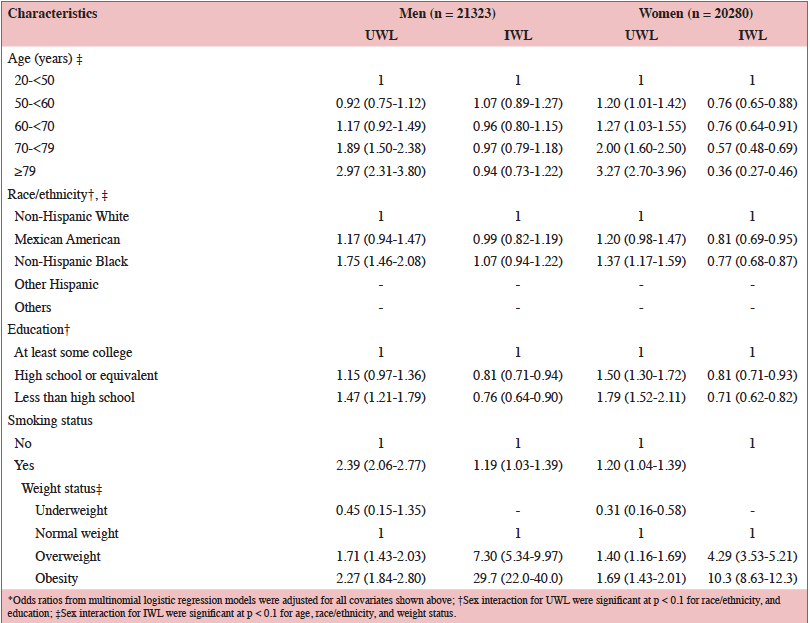
Life expectancies in European Union (EU) countries continue to rise, expanding the aging population. It has been estimated that by 2050 greater than 25% of the EU population will be ≥65 years of age, which has the potential to challenge the healthcare system (1). According to the European Health Report, important non-communicable diseases (NCDs) associated with aging include cardiovascular disease (CVD), certain cancers, and type 2 diabetes (1). Because these NCDs are the leading causes of early mortality in the EU, primary and secondary prevention efforts are needed to reduce NCD-related morbidity and mortality (1). To allow older individuals to live more independently and remain integrated in society, the World Health Organization’s agenda for preventing NCDs includes increasing physical activity, reducing tobacco and alcohol use, and reducing the risk of malnutrition, a condition in which many nutrient requirements are not met (2). The European Food Safety Authority (EFSA) reference intakes for selected micronutrients in adults >50 years of age are listed in Table 1 (3).

Table 1. European Food Safety Authority (EFSA) daily reference intakes for selected micronutrients in adults >50 years of age (3)

Table 1. European Food Safety Authority (EFSA) daily reference intakes for selected micronutrients in adults >50 years of age (3)

*1 μg of DFE equals 1 μg of food folate=0.6 μg of folic acid from fortified food=0.5 μg of a folic acid supplement; †1 μg RE equals 1 μg of retinol, 6 μg of β-carotene, and 12 μg of other provitamin A carotenoids; DFE, dietary folate equivalent; RE, retinol equivalent.


According to a systematic review of longitudinal data, risk factors for malnutrition include frailty, excessive polypharmacy, declining physical functioning and cognition, depression, dysphagia, and institutionalization (4). Multiple physical, socioeconomic, and cultural factors affect the nutritional status of older individuals, including changes in the ability to absorb nutrients, reduced appetite, and decreased ability to chew (4, 5). Furthermore, there are multiple drug-nutrient interactions that should be considered for this population (Table 2) (6). Insufficient energy intake associated with aging is complex and may involve chronic illnesses and reduced ability and desire to prepare and eat meals (7). Micronutrient intake below recommended amounts increases NCD risks (8). For example, micronutrient deficiencies can cause mitochondrial decay, a mechanism contributing to aging and development of diseases including cancer and neural decay (8). A relationship has been observed between the intake of certain micronutrients (i.e. vitamins D, B6, B12, and E and folate) and frailty in older adults (9). Values for ranges of nutrient intakes in the EU are described in Table 3 (10).

Table 2. Common drug-micronutrient interactions and consequences of the interaction(s) (6)

Table 2. Common drug-micronutrient interactions and consequences of the interaction(s) (6)

Table 3. Nutrient intake in the European Union based on national data (10)

Table 3. Nutrient intake in the European Union based on national data (10)

*Intakes reported for individuals 19–64 years of age for all countries except the following: Greece: 22±2 years of age; Hungary: ≥18 years of age; United Kingdom: 25–64 years of age; †Intakes reported for individuals >64 years of age for all countries except Hungary (>59 years of age); ‡Folate equivalent; 1 μg food folate=0.5 μg folic acid (PGA)=0.6 μg folic acid taken with meals; §RRR-α-tocopherol equivalent=mg α-tocopherol + mg β-tocopherol x 0.5 + mg y-tocopherol x 0.25 + mg α-tocotrienol x 0.33. DFE, dietary folate equivalent; PGA, pteroyl glutamic acid.


This narrative review describes the nutritional needs of middle-aged (50–64 years) and older (≥65 years) adults in the EU and interventions healthcare professionals should consider. Literature searches were conducted to identify sources that evaluated the nutritional status and interventions relevant to this population, with an emphasis placed on systematic reviews, meta-analyses, and dietary guidelines. Notably, some meta-analyses also included other ages (<50 years), but those that are included primarily assessed older individuals.


Protein

Aging leads to a loss of muscle mass and strength and poor physical performance (i.e. sarcopenia) that has been associated with macro- and micronutrient deficiencies, suggesting that a high-quality diet that includes optimal protein and nutrient intake combined with physical exercise can reduce this risk (11). Sarcopenia can increase the risk for falls, fractures, disability, loss of independence, and increased mortality (11). The prevalence of sarcopenia is approximately 1–29% in community-dwelling older adults and 10–33% in those living in long-term care and acute hospital settings (12).
Adequate protein intake is important to healthy aging, and higher intakes may be necessary to compensate for the difficulty of maintaining muscle mass; however, recommendations vary by country (13). The EFSA recommendation for protein intake for adults is 0.83 g/kg/d (3), yet, the PROT-AGE Study Group recommends protein intakes in older adults of 1.0–1.2 g/kg/d (13). The authors state that those with chronic diseases, severe illnesses, injury, or malnutrition may require higher intakes (i.e. 1.2–1.5 and 2.0 g/kg/d, respectively) (13). Higher protein intake can negatively impact kidney function in those with severe kidney disease not receiving dialysis; therefore, caution should be taken in this population (13). The Nordic Nutrition Recommendations set a tentative recommendation of 1.2–1.5 g/kg/d while stressing that adequate data do not exist to estimate an optimal protein intake (14).
Protein quality, timing of administration, whether to supplement with single amino acids, and the addition of physical exercise should be considered (13). Protein supplementation immediately following resistance training exercise is beneficial for muscle mass and strength (13). A recent meta-analysis of randomized, controlled trials (RCTs) conducted with whey-, leucine-, and casein-based protein supplements combined with resistance training reported that elderly individuals adhering to this regimen improved lean body and appendicular mass, body fat and mass, muscle strength, and mobility (15). However, the International Sarcopenia Initiative stated that protein supplements alone or combined with resistance training have shown inconsistent effects on muscle mass and function in individuals ≥50 years of age (12).


Dietary fiber

Dietary fiber is critical to maintaining proper laxation and a healthy microbiome and is involved in many physiological activities including protecting against CVD (5). EFSA recommends that older adults consume 25 g/d of total fiber (3), but few meet this goal (5). The European Society of Cardiology (ESC) and European Atherosclerosis Society (EAS) recommendations for managing dyslipidemia include consuming 45% to 55% of energy from carbohydrates, including fruit, vegetables, whole grains, legumes, and nuts, and 25-40 grams of total dietary fiber, such as β-glucan from oat and barley (16).
A meta-analysis of 67 RCTs reported that consuming high-fiber diets significantly reduced total and low-density lipoprotein (LDL) cholesterol, but not high-density lipoprotein cholesterol (17). Other meta-analyses of RCTs reported that using different fiber supplements significantly reduced diastolic blood pressure, an effect that was more pronounced in adults >40 years of age compared with younger adults (18), and significantly reduced glycated hemoglobin in middle-aged and older adults with type 2 diabetes (19).


Omega-3 fatty acids

Omega-3 fatty acids have anti-inflammatory properties, and higher intakes have been linked to reductions in CVD risk and cognitive impairment (5). Omega-3 fatty acids cannot be efficiently converted from dietary alpha-linolenic acid in the body; therefore, docosahexaenoic acid (DHA) and eicosapentaenoic acid (EPA) often do not reach adequate levels. Dietary sources of omega-3 fatty acids include oily fish, flaxseed, and walnuts (5). EFSA recommends that adults consume 0.5% of their energy in the form of alpha-linolenic acid and 250 mg/d EPA plus DHA (3). ESC and EAS recommendations include 2–3 g/d of long-chain omega-3 fatty acid supplements to reduce triglycerides and regular consumption of fish and nuts for preventing CVD (20).
Meta-analyses of RCTs conducted in adults with or without cardiovascular comorbidities (e.g. coronary heart disease, heart failure) receiving DHA and EPA through the diet or supplementation reported a greater risk reduction among individuals with elevated triglyceride and LDL cholesterol levels (21) and significantly improved brain natriuretic peptide and serum norepinephrine levels (22). The Multicenter Osteoarthritis Study, a prospective cohort study of individuals (mean age: 60 years) with, or at high risk for, knee osteoarthritis (OA) reported that individuals with high plasma omega-3 fatty acid levels, especially DHA, had less patellofemoral cartilage loss (23). Well-controlled RCTs are necessary to support using omega-3 fatty acid supplements for OA.
Due to the association between inadequacies in omega-3 fatty acid status and cognitive decline, a number of studies have been conducted to evaluate the effects of omega-3 fatty acids on cognitive functioning. Meta-analyses of observational studies and RCTs conducted in middle-aged and older adults reported significant improvements in cognitive functioning (i.e. attention, executive functioning, memory), but no improvements in other cognitive parameters were observed (24, 25). A more recent meta-analysis of omega-3 fatty acid trials in adults (>40 years of age) did not observe improvements in cognitive functioning (26).


Folate and vitamins B6 and B12

Folate is a B-vitamin involved in the metabolism of nucleic acid precursors, DNA methylation, homocysteine metabolism, and cognition (5). Vitamin B6 is essential for many enzymatic reactions involved in protein metabolism, while vitamin B12 is involved in folate metabolism and neurological functioning. Deficiencies in vitamin B12 can cause peripheral neuropathy and cognitive dysfunction (3, 5). EFSA recommends that adults consume 250 μg/d folate, 4.0 μg/d vitamin B12, and 1.5 mg/d and 1.3 mg/d vitamin B6, in men and women, respectively (3). Deficiencies and inadequacies in folate and vitamins B6 and B12 can increase homocysteine concentrations, weaken immunity, and increase risk of CVD, stroke, cognitive dysfunction, depression, osteoporosis, and fracture risk (5, 27). In the EU up to 20% of older adults (>64 years) do not obtain adequate amounts of vitamin B12, and a substantial proportion (17–46%) do not achieve adequate intake of folate (28). Older adults may have difficulty extracting vitamin B12 from natural food sources due to an age-related decline in gastric acid secretion (5), which reduces the ability of vitamin B12 to bind to intrinsic factor (29).
Meta-analyses of folic acid supplement RCTs in middle-aged and older adults reported significant risk reductions in stroke and CVD-related events (30) and significant reductions in plasma homocysteine concentration, with further reductions when vitamin B12 was co-administered (31).
Folic acid and vitamin B12 supplementation may also improve bone health, primarily due to the effects on homocysteine levels (32). However, an RCT conducted in older individuals (≥65 years) with elevated homocysteine levels supplemented with folic acid and vitamin B12 reported no reduction in fracture risk, aside from a sub-group of individuals >80 years of age (32).
A meta-analysis of case-control studies reported significant associations between higher folate intake and decreased risk of head and neck squamous cell carcinoma (33). Despite these potential benefits, excessive intake of synthetic folic acid can increase the risk of certain cancers (34). Therefore, it is important to determine an individual’s intake of dietary folate and folic acid from supplements and fortification to avoid reaching levels that can cause adverse health outcomes.
In meta-analyses of RCTs of folic acid in subjects ≥45 years of age without dementia (35) and vitamin B6 and B12 in subjects ≥40 years of age who were healthy or at risk for CVD (26), no improvements in cognitive functioning were found. Another trial that administered folic acid and vitamin B12 to older adults (60–74 years) reported significant improvements in overall cognitive functioning and immediate and delayed recall (36).


Vitamin A

Vitamin A is a fat-soluble vitamin found either as preformed vitamin A (retinol) in animal products such as liver and dairy, or as provitamin A carotenoids such as β-carotene in fruit and vegetables (3, 37). Vitamin A is involved in regulating cellular growth and differentiation and is required for healthy immune function and vision (3, 37). EFSA recommends that adult men and women consume 750 μg/d and 650 μg/d vitamin A (as Retinol Equivalents, Table 1), respectively, from a mixture of preformed vitamin A and provitamin A carotenoids (3).
A critical issue with vitamin A is its toxicity (hypervitaminosis A) caused by excessive retinol intake; middle-aged and older adults may be particularly susceptible to vitamin A toxicity (38). Additionally, several prospective cohort studies have reported that higher intakes of retinol are associated with an increased risk of hip fractures in primarily middle-aged and older adults (39), and two RCTs found that high-dose supplementation with β-carotene (alone or combined with retinol) further increased the risk of lung cancer in at-risk populations (e.g. smokers, asbestos-exposed workers) (38). However, a meta-analysis of four RCTs found no effect on lung cancer risk with retinol or β-carotene supplementation in healthy adults (40).


Vitamin D

Vitamin D plays critical roles in calcium metabolism and bone health but is also involved in other health outcomes, such as neurological conditions, autoimmune diseases, NCDs (e.g. type 2 diabetes, cancers), and infections (5, 41). EFSA recommends vitamin D intake of 15 µg/d for adults (3), while the International Osteoporosis Foundation recommends average daily intake of 20–25 µg/d for older adults (41). Older adults may be at particular risk for insufficiency and deficiency due to lower sunlight exposure, reduced ability of the skin to synthesize vitamin D from 7-dehydrocholesterol upon sunlight exposure, and limited consumption of food sources of vitamin D (e.g. fortified milk, oily fish) (5). Across the EU, the percentage of the older population (>64 years) not obtaining adequate amounts of vitamin D is approximately 90% in most areas except Norway, Finland, and Spain (28). A systematic review of 195 studies reported that mean 25(OH)D values for those >65 years of age in the EU were 51.7 nmol/L (42). European League Against Rheumatism (EULAR) and European Federation of National Associations of Orthopaedics and Traumatology (EFORT) recommendations for preventing future fractures in those ≥50 years of age with previous fractures taking anti-osteoporosis drugs include supplementation with 800 IU/d (equivalent to
20 µg/d) vitamin D with adequate calcium intake (1000–1200 mg/d) (43). This recommendation is based on a meta-analysis of RCTs demonstrating a significant reduction in the risk of falling with 700–1000 IU/d (17.5–25 µg/d) vitamin D or 25(OH)D levels of 60–95 nmol/L (44) and a pooled analysis demonstrating a reduced risk of hip fractures with ≥800 IU/d (≥20 µg/d) vitamin D or baseline 25(OH)D levels >60 nmol/L in elderly individuals (≥65 years) (45). A separate meta-analysis of RCTs reported that vitamin D at 482–770 IU/d (12–19 µg/d) reduced the risk for non-vertebral and hip fractures by 20% (44), while a National Osteoporosis Foundation meta-analysis of RCTs reported that vitamin D supplements of 400–800 IU/d (10–20 µg/d) and calcium of 500–1200 mg/d reduced the risk of total and hip fractures by 15% and 30%, respectively (46). Evidence for the effectiveness of vitamin D supplements in reducing the risk of falling is inconsistent, and bolus doses have been shown to increase the risk for falling (47).
It has been suggested that vitamin D can have positive effects on other health outcomes, including autoimmune diseases and CVD, type 2 diabetes, and cancer (48). Deficiencies in 25(OH)D levels may also be associated with increased risk of colorectal and breast cancer, cardiovascular events, and mortality (48). Aggregated evidence from RCTs suggests that vitamin D supplementation could effectively prevent respiratory tract infections (49, 50), which caused 2.8 million deaths worldwide in 2010 (49) and affect the elderly at increased rates (50).


Calcium

Calcium is an essential mineral that is involved in promoting bone health but is also associated with other health outcomes such as controlling blood pressure (5). While EFSA recommends 750 mg/d total calcium intake in adults >50 years of age (3), EULAR/EFORT recommends 1000–1200 mg/d for preventing fractures in those using anti-osteoporosis drugs, with supplementation as necessary (43). EULAR/EFORT also cautions that calcium supplements may produce adverse gastrointestinal and possibly cardiovascular effects (43). Across the EU, the percentage of the older population (>64 years of age) not obtaining adequate amounts of calcium ranges from 48–100% (28).
A meta-analysis of 29 RCTs that evaluated calcium supplementation with or without vitamin D on bone health outcomes in middle-aged and older individuals (≥50 years of age) observed significant reductions in fracture risk and bone loss (51). Concern has been raised about the potential for calcium supplements to increase the risk for CVD, but a long-term study specifically designed to evaluate this potential found no evidence of increased risk in older women (mean age: 75 years) in relation to placebo (52). Another meta-analysis reported that total calcium intake (diet and supplementation) below the tolerable upper intake level (UL) is not associated with an increased risk for CVD (53). However, caution should be taken when recommending calcium supplementation to those already obtaining adequate dietary intake (53).


Vitamin K

Vitamin K describes a group of related fat-soluble vitamins critically involved in coagulation and bone health by activating specific proteins in the bloodstream and bone (3, 5, 54). Vitamin K1 (phylloquinone) is the primary dietary form of vitamin K, which is found in green leafy vegetables and vegetable oils, while vitamin K2 (menaquinone) is primarily found in animal-based or fermented foods (3, 54). EFSA recommends that adults consume 70 µg/d vitamin K (3). The mechanism of action for a class of anticoagulant therapies (e.g. warfarin) used for preventing atrial fibrillation and other cardiovascular events involves vitamin K antagonism; therefore, balancing the dose of these treatments and dietary vitamin K intake should be considered (55). However, studies have provided conflicting results; some show a negative relationship between coagulation stability and vitamin K intake and others suggest that some amount of vitamin K intake is necessary to produce an adequate response (55). A Cochrane database review reported that only one study showed this additive anticoagulant effect, indicating that the current evidence is insufficient to recommend vitamin K for those with unstable response to warfarin (56). Supplemental vitamin K has a strong anticoagulant effect, and consuming high levels of vitamin K-rich foods may interact with anticoagulant treatment (57). Vitamin K1 supplementation combined with calcium and vitamin D3 has also been shown to modestly improve bone mineral content in older non-osteoporotic women; however, these effects were not observed with vitamin K1 alone, suggesting that there may be a synergistic effect of these nutrients (58). Vitamin K2 has also been shown to produce benefits in arterial stiffness and bone mineral density (59, 60).


Vitamin E

Vitamin E (α-tocopherol) is a fat-soluble vitamin with antioxidant properties that are critical for protecting polyunsaturated fatty acids in membrane phospholipids and plasma lipoproteins from oxidative damage (3); it is also involved in immune function (5). α-Tocopherol deficiency causes the development of neurological symptoms (e.g. ataxia) (3). EFSA recommends that adult males and females consume 13 mg/d and 11 mg/d vitamin E, respectively (3), yet those >64 years of age have been shown to consume only between 6.3 and 13.7 mg/d (10). Very few individuals meet intake recommendations for vitamin E through diet alone (5). A meta-analysis of dietary intake studies reported that vitamin E is associated with a dose-dependent reduction in lung cancer risk (61), but another meta-analysis reported no effect on total or cancer-related mortality, aside from a significant reduction in the incidence of prostate cancer when vitamin E was consumed with other nutrients (62). Another meta-analysis of RCTs reported a decreased risk of ischemic stroke but an increased risk for hemorrhagic stroke. Notably, the doses of vitamin E administered substantially exceeded recommended intake levels (63).


Vitamin C

Vitamin C is a water-soluble vitamin with strong reducing and antioxidant properties; it acts as a cofactor in several enzymatic reactions for the synthesis of carnitine, catecholamines, and pro-collagen and for metabolizing cholesterol to bile acids (3). The primary dietary sources of vitamin C include fruit and vegetables and their juices (3). EFSA recommends that adult males and females consume 90 mg/d and 80 mg/d vitamin C, respectively (3). In the EU, the proportion of individuals >64 years of age falling below the average requirement of vitamin C is 4-33% (28).
Population-based studies have shown that plasma vitamin C levels are significantly and inversely related to stroke risk (64). Furthermore, lower vitamin C levels have been linked with a greater risk for Alzheimer’s disease (65, 66). Meta-analyses of RCTs have found that vitamin C supplementation significantly reduces both systolic and diastolic blood pressure (67), improves endothelial function and vasodilation in individuals with cardiometabolic risk factors (68), and decreases risk for lung cancer (69) and age-related cataracts (70).


Magnesium

Magnesium is an essential mineral for many enzymatic reactions involved in the synthesis of carbohydrates, lipids, nucleic acids, and proteins, and serves in various neurological and cardiovascular functions, including regulating blood pressure (3, 5). Magnesium is primarily found in muscle tissues and is an important component of bone (3, 5). Magnesium occurs naturally in a number of food items, including nuts, whole grains, seafood, fruit, and vegetables (3). EFSA recommends that adult men and women consume 350 mg/d and 300 mg/d of magnesium, respectively (3). There are few clear indications of magnesium inadequacies due to its various metabolic effects, and serum magnesium concentration as an indicator of status is questionable since there are no reliable biomarkers for magnesium body status (3).
Meta-analyses of prospective cohort and observational studies conducted primarily in middle-aged individuals have reported an association between higher circulating magnesium concentrations and decreased CVD risk (71), a significant inverse relationship between dietary magnesium intake and risk of metabolic syndrome (72), and a relationship between higher magnesium intake and reductions in colorectal cancer (73). Meta-analyses of RCTs that evaluated magnesium supplementation on diabetes-related outcomes reported improvements in insulin resistance (74) and fasting glucose levels (75) and significant reductions in systolic and diastolic blood pressure (76), including in individuals with insulin resistance, pre-diabetes, and other NCDs (77).


Potassium

Potassium is the primary intracellular cation responsible for maintaining fluid and electrolyte balance in the body and, hence, proper nerve conduction, muscle contraction, blood volume, and blood pressure (3, 5). Potassium occurs naturally in all foods but is particularly represented in root vegetables, fruit, whole grains, coffee, and dairy (3). EFSA recommends that adults consume 3500 mg/d of potassium, but these values can vary by country (3). Insufficient potassium intake causes hypertension and increases the risk of CVD, kidney stones, and osteoporosis (5).
A meta-analysis of prospective cohort studies reported that higher dietary potassium intake reduced the risk of stroke, which the authors attributed to reduced blood pressure (78). Although a 2006 Cochrane database review did not find substantial support for potassium supplementation for hypertension (79), a more recent meta-analysis of RCTs reported that potassium supplementation reduced systolic and diastolic blood pressure in a dose-dependent manner in hypertensive individuals (80).


Ensuring adequate nutrition in middle-aged and older adults

There is a robust body of evidence suggesting that a whole-diet approach not only lowers mortality from NCDs, but also positively impacts physical and cognitive functioning, mental health, and quality of life in older adults (81). However, too few high-quality studies have evaluated these outcomes to make clear recommendations. In addition to the nutritional benefits of a healthful diet, there are psychological benefits to eating that should not be overlooked (82). Increasing contact with professionals, identifying barriers, developing problem-solving techniques, and setting appropriate goals can improve the outcomes of dietary interventions in middle-aged and older individuals (83). Nutrition education programs have been shown to be effective for improving the nutritional status of older adults, particularly when they include specific interventions or multiple sessions (84).
Higher adherence to a Mediterranean Diet has been found to be significantly and inversely related to overall mortality in adults >65 years of age (85) and to reduced all-cause, CVD- and cancer-related mortality (86). Furthermore, a multivitamin/multimineral supplement (MVMS) that provides most micronutrients in recommended amounts can provide nutritional support for those who are unable to reach adequate micronutrient intake levels from their habitual diet, particularly for older individuals who often experience malnutrition with advancing age (87).
Healthcare providers commonly recommend MVMS to older adults, and while MVMS are generally safe (88), their benefits for improving health-related outcomes have been difficult to conclusively demonstrate (89). The Physicians’ Health Study II (PHS II) observed no reduction in the risk for developing CVD (90), but there was a significant 8% reduction in the risk of all types of cancer in this middle-aged and older male population (≥50 years of age) taking a daily MVMS for a mean duration of 11 years (91). The reduction in cancer risk was 12% when excluding prostate cancer from the analysis, and even greater (27%) in men with a baseline history of cancer (91). Despite its long duration and large sample size involving more than 14,000 male physicians, PHS II was insufficiently powered to detect statistically significant effects of MVMS on any individual type of cancer (91).
PHS II also found a significant 9% reduction in total age-related cataracts and an 11% reduction in cataract surgery (92). According to a Cochrane database review, use of an MVMS with antioxidant vitamins and minerals may delay the progression of age-related macular degeneration (AMD) (93). Lutein and zeaxanthin, which are sometimes added to MVMS formulations, also seem to be beneficial for the management of AMD (94). A meta-analysis of eight RCTs utilizing lutein and zeaxanthin supplementation showed improvements in visual acuity and contrast sensitivity in subjects with AMD (95).
Conflicting evidence on cognition has been observed in older adults who use MVMS. One clinical trial in middle-aged and older men reported improvements in episodic memory, including contextual recognition (96), while another trial in healthy middle-aged and older adults reported no benefit in cognitive task performance (97). Both of these studies reported that MVMS use improved some health-related biomarkers (i.e. C-reactive protein, liver function, and vitamin B6 and B12 blood levels, cholesterol, and homocysteine levels) (96, 97). A meta-analysis of 10 RCTs in primarily middle-aged and older adults reported that MVMS modestly improved some aspects of memory (98). An MVMS formulated with folic acid and vitamins B6 and B12 was shown to improve plasma homocysteine concentrations in an RCT in individuals ≥50 years of age (99), and another study in older individuals being treated with metformin demonstrated that use of an MVMS can reduce the risk for metformin-mediated deficiencies in vitamin B12 levels (100).
It is important that an MVMS include nutrients only in amounts that approximate reference intake values, and middle-aged and older adults who decide to use an MVMS should be aware that using additional single-nutrient supplements could result in a total intake exceeding the UL of these nutrients, increasing the risk of adverse health outcomes (87). Furthermore, there may be some individual exceptions to consider for these populations. For example, the typical dose of calcium commonly used in an MVMS may not be sufficient to promote bone health; therefore, dietary intake should be considered (89).


Conclusion

Data from RCTs suggest that dietary interventions and, when warranted, supplementation with MVMS can be used to reduce the risk of experiencing nutritional deficiencies and inadequacies that are detrimental to the health of middle-aged and older adults. For individuals who do not consume adequate protein, fiber, omega-3 fatty acids, and micronutrients from their habitual diet, supplementation may be required. The individual’s medical history, medication use, dietary patterns, and other risk factors should be considered when recommending dietary supplements.


Funding
Medical writing support was provided by Dennis Stancavish of Peloton Advantage, LLC, and was funded by Pfizer.

Role of the Sponsor
The sponsor was involved in the review and approval of the manuscript.

Conflicts of Interest
Susanne Bügel has received research grants from the Software AG Foundation and ERASMUS+. She is a board member of Food, Quality and Health and the Federation of European Nutrition Societies and as such receives travel support for meetings supported by these organizations. Balz Frei is the past recipient of multiple grants from the US National Institutes of Health and currently serves as a consultant for Pfizer Consumer Healthcare and DSM Nutritional Products.


References

1. World Health Organization. The European health report 2012: Charting the way to well-being. 2013. World Health Organization, Geneva, Switzerland.
2. World Health Organization Regional Office for Europe. Strategy and action plan for healthy ageing in Europe, 2012-2020. 2012. World Health Organization Regional Office for Europe, Copenhagen, Denmark.
3. Online document. European Food Safety Authority. Dietary reference values for nutrients, summary report. European Food Safety Authority. 2017. https://www.efsa.europa.eu/sites/default/files/2017_09_DRVs_summary_report.pdf. Accessed November 21, 2017.
4. Favaro-Moreira NC, Krausch-Hofmann S, Matthys C, et al. Risk factors for malnutrition in older adults: a systematic review of the literature based on longitudinal data. Adv Nutr 2016;7:507-522.
5. National Academies of Sciences Engineering Medicine. Meeting the dietary needs of older adults: Exploring the impact of the physical, social, and cultural environment: Workshop summary. 2016. National Academies Press, Washington, DC.
6. Higdon J, Drake VJ. Drug-nutrient interactions. 2012. Georg Thieme Verlag, Stuttgart, Germany.
7. ter Borg S, Verlaan S, Hemsworth J, et al. Micronutrient intakes and potential inadequacies of community-dwelling older adults: a systematic review. Br J Nutr 2015;113:1195-1206.
8. Ames BN. Low micronutrient intake may accelerate the degenerative diseases of aging through allocation of scarce micronutrients by triage. Proc Natl Acad Sci U S A 2006;103:17589-17594.
9. Lorenzo-Lopez L, Maseda A, de Labra C, Regueiro-Folgueira L, Rodriguez-Villamil JL, Millan-Calenti JC. Nutritional determinants of frailty in older adults: A systematic review. BMC Geriatr 2017;17:108.
10. Elmadfa I, Meyer A, Nowak V, et al. European nutrition and health report 2009. Forum Nutr 2009;62:1-405.
11. Nowson C, O’Connell S. Protein requirements and recommendations for older people: A review. Nutrients 2015;7:6874-6899.
12. Cruz-Jentoft AJ, Landi F, Schneider SM, et al. Prevalence of and interventions for sarcopenia in ageing adults: a systematic review. Report of the International Sarcopenia Initiative (EWGSOP and IWGS). Age Ageing 2014;43:748-759.
13. Bauer J, Biolo G, Cederholm T, et al. Evidence-based recommendations for optimal dietary protein intake in older people: a position paper from the PROT-AGE Study Group. J Am Med Dir Assoc 2013;14:542-559.
14. Pedersen AN, Cederholm T. Health effects of protein intake in healthy elderly populations: a systematic literature review. Food Nutr Res 2014;58.
15. Liao CD, Tsauo JY, Wu YT, et al. Effects of protein supplementation combined with resistance exercise on body composition and physical function in older adults: a systematic review and meta-analysis. Am J Clin Nutr 2017;106:1078-1091.
16. Catapano AL, Graham I, De Backer G, et al. 2016 ESC/EAS guidelines for the management of dyslipidaemias. Eur Heart J 2016;37:2999-3058.
17. Brown L, Rosner B, Willett WW, Sacks FM. Cholesterol-lowering effects of dietary fiber: a meta-analysis. Am J Clin Nutr 1999;69:30-42.
18. Streppel MT, Arends LR, van ‘t Veer P, Grobbee DE, Geleijnse JM. Dietary fiber and blood pressure: a meta-analysis of randomized placebo-controlled trials. Arch Intern Med 2005;165:150-156.
19. Post RE, Mainous AG 3rd, King DE, Simpson KN. Dietary fiber for the treatment of type 2 diabetes mellitus: a meta-analysis. J Am Board Fam Med 2012;25:16-23.
20. Reiner Z, Catapano AL, De Backer G, et al. ESC/EAS Guidelines for the management of dyslipidaemias: the Task Force for the management of dyslipidaemias of the European Society of Cardiology (ESC) and the European Atherosclerosis Society (EAS). Eur Heart J 2011;32:1769-1818.
21. Alexander DD, Miller PE, Van Elswyk ME, Kuratko CN, Bylsma LC. A meta-analysis of randomized controlled trials and prospective cohort studies of eicosapentaenoic and docosahexaenoic long-chain omega-3 fatty acids and coronary heart disease risk. Mayo Clin Proc 2017;92:15-29.
22. Wang C, Xiong B, Huang J. The role of omega-3 polyunsaturated fatty acids in heart failure: a meta-analysis of randomised controlled trials. Nutrients 2016;9.
23. Baker KR, Matthan NR, Lichtenstein AH, et al. Association of plasma n-6 and n-3 polyunsaturated fatty acids with synovitis in the knee: the MOST study. Osteoarthritis Cartilage 2012;20:382-387.
24. Jiao J, Li Q, Chu J, Zeng W, Yang M, Zhu S. Effect of n-3 PUFA supplementation on cognitive function throughout the life span from infancy to old age: a systematic review and meta-analysis of randomized controlled trials. Am J Clin Nutr 2014;100:1422-1436.
25. Yurko-Mauro K, Alexander DD, Van Elswyk ME. Docosahexaenoic acid and adult memory: a systematic review and meta-analysis. PLoS One 2015;10:e0120391.
26. Forbes SC, Holroyd-Leduc JM, Poulin MJ, Hogan DB. Effect of nutrients, dietary supplements and vitamins on cognition: a systematic review and meta-analysis of randomized controlled trials. Can Geriatr J 2015;18:231-245.
27. Porter K, Hoey L, Hughes CF, Ward M, McNulty H. Causes, consequences and public health implications of low B-vitamin status in ageing. Nutrients 2016;8:725.
28. Roman Viñas B, Barba LR, Ngo J, et al. Projected prevalence of inadequate nutrient intakes in Europe. Ann Nutr Metab 2011;59:84-95.
29. Russell RM. Factors in aging that effect the bioavailability of nutrients. J Nutr 2001;131:1359s-1361s.
30. Li Y, Huang T, Zheng Y, Muka T, Troup J, Hu FB. Folic acid supplementation and the risk of cardiovascular diseases: a meta-analysis of randomized controlled trials. J Am Heart Assoc 2016;5.
31. Homocysteine Lowering Trialists’ Collaboration Dose-dependent effects of folic acid on blood concentrations of homocysteine: a meta-analysis of the randomized trials. Am J Clin Nutr 2005;82:806-812.
32. van Wijngaarden JP, Swart KM, Enneman AW, et al. Effect of daily vitamin B-12 and folic acid supplementation on fracture incidence in elderly individuals with an elevated plasma homocysteine concentration: B-PROOF, a randomized controlled trial. Am J Clin Nutr 2014;100:1578-1586.
33. Fan C, Yu S, Zhang S, Ding X, Su J, Cheng Z. Association between folate intake and risk of head and neck squamous cell carcinoma: An overall and dose-response PRISMA meta-analysis. Medicine (Baltimore) 2017;96:e8182.
34. Cole BF, Baron JA, Sandler RS, et al. Folic acid for the prevention of colorectal adenomas: a randomized clinical trial. JAMA 2007;297:2351-2359.
35. Wald DS, Kasturiratne A, Simmonds M. Effect of folic acid, with or without other B vitamins, on cognitive decline: meta-analysis of randomized trials. Am J Med 2010;123:522-527.
36. Walker JG, Batterham PJ, Mackinnon AJ, et al. Oral folic acid and vitamin B-12 supplementation to prevent cognitive decline in community-dwelling older adults with depressive symptoms–the Beyond Ageing Project: a randomized controlled trial. Am J Clin Nutr 2012;95:194-203.
37. Sherwin JC, Reacher MH, Dean WH, Ngondi J. Epidemiology of vitamin A deficiency and xerophthalmia in at-risk populations. Trans R Soc Trop Med Hyg 2012;106:205-214.
38. Russell RM. The vitamin A spectrum: from deficiency to toxicity. Am J Clin Nutr 2000;71:878-884.
39. Wu AM, Huang CQ, Lin ZK, et al. The relationship between vitamin A and risk of fracture: meta-analysis of prospective studies. J Bone Miner Res 2014;29:2032-2039.
40. Cortes-Jofre M, Rueda JR, Corsini-Munoz G, Fonseca-Cortes C, Caraballoso M, Bonfill Cosp X. Drugs for preventing lung cancer in healthy people. Cochrane Database Syst Rev 2012;10:CD002141.
41. Dawson-Hughes B, Mithal A, Bonjour JP, et al. IOF position statement: vitamin D recommendations for older adults. Osteoporos Int 2010;21:1151-1154.
42. Hilger J, Friedel A, Herr R, et al. A systematic review of vitamin D status in populations worldwide. Br J Nutr 2014;111:23-45.
43. Lems WF, Dreinhofer KE, Bischoff-Ferrari H, et al. EULAR/EFORT recommendations for management of patients older than 50 years with a fragility fracture and prevention of subsequent fractures. Ann Rheum Dis 2017;76:802-810.
44. Bischoff-Ferrari HA, Willett WC, Wong JB, et al. Prevention of nonvertebral fractures with oral vitamin D and dose dependency: a meta-analysis of randomized controlled trials. Arch Intern Med 2009;169:551-561.
45. Bischoff-Ferrari HA, Willett WC, Orav EJ, et al. A pooled analysis of vitamin D dose requirements for fracture prevention. N Engl J Med 2012;367:40-49.
46. Weaver CM, Alexander DD, Boushey CJ, et al. Calcium plus vitamin D supplementation and risk of fractures: an updated meta-analysis from the National Osteoporosis Foundation [published erratum and additional analyses appear in Osteoporos Int. 2016;27(8):2643-2646] Osteoporos Int 2016;27:367-376.
47. Gallagher JC. Vitamin D and falls – the dosage conundrum. Nat Rev Endocrinol 2016;12:680-684.
48. Bruyere O, Cavalier E, Souberbielle JC, et al. Effects of vitamin D in the elderly population: current status and perspectives. Arch Belg Sante Publ 2014;72:32.
49. Bergman P, Lindh AU, Bjorkhem-Bergman L, Lindh JD. Vitamin D and respiratory tract infections: A systematic review and meta-analysis of randomized controlled trials. PLoS One 2013;8:e65835.
50. Katona P, Katona-Apte J. The interaction between nutrition and infection. Clin Infect Dis 2008;46:1582-1588.
51. Tang BM, Eslick GD, Nowson C, Smith C, Bensoussan. A Use of calcium or calcium in combination with vitamin D supplementation to prevent fractures and bone loss in people aged 50 years and older: a meta-analysis. Lancet 2007;370:657-666.
52. Lewis JR, Calver J, Zhu K, Flicker L, Prince RL. Calcium supplementation and the risks of atherosclerotic vascular disease in older women: results of a 5-year RCT and a 4.5-year follow-up. J Bone Miner Res 2011;26:35-41.
53. Chung M, Tang AM, Fu Z, Wang DD, Newberry SJ. Calcium intake and cardiovascular disease risk: An updated systematic review and meta-analysis. Ann Intern Med 2016;165:856-866.
54. Grober U, Reichrath J, Holick MF, Kisters K Vitamin K: an old vitamin in a new perspective. Dermatoendocrinol 2014;6:e968490.
55. Violi F, Lip GY, Pignatelli P, Pastori D Interaction between dietary vitamin K intake and anticoagulation by vitamin K antagonists: Is it really true?: a systematic review. Medicine (Baltimore) 2016;95:e2895.
56. Mahtani KR, Heneghan CJ, Nunan D, Roberts NW. Vitamin K for improved anticoagulation control in patients receiving warfarin. Cochrane Database Syst Rev 2014;5:CD009917.
57. Bugel SG, Spagner C, Poulsen SK, Jakobsen J, Astrup A. Phylloquinone content from wild green vegetables may contribute substantially to dietary intake. Can J Agric Crops 2016;1:83-88.
58. Bolton-Smith C, McMurdo ME, Paterson CR, et al. Two-year randomized controlled trial of vitamin K1 (phylloquinone) and vitamin D3 plus calcium on the bone health of older women. J Bone Miner Res 2007;22:509-519.
59. Knapen MH, Drummen NE, Smit E, Vermeer C, Theuwissen E. Three-year low-dose menaquinone-7 supplementation helps decrease bone loss in healthy postmenopausal women. Osteoporos Int 2013;24:2499-2507.
60. Knapen MH, Braam LA, Drummen NE, Bekers O, Hoeks AP, Vermeer C. Menaquinone-7 supplementation improves arterial stiffness in healthy postmenopausal women. A double-blind randomised clinical trial. Thromb Haemost 2015;113:1135-1144.
61. Zhu YJ, Bo YC, Liu XX, Qiu CG. Association of dietary vitamin E intake with risk of lung cancer: a dose-response meta-analysis. Asia Pac J Clin Nutr 2017;26:271-277.
62. Alkhenizan A, Hafez K. The role of vitamin E in the prevention of cancer: a meta-analysis of randomized controlled trials. Ann Saudi Med 2007;27:409-414.
63. Schurks M, Glynn RJ, Rist PM, Tzourio C, Kurth T. Effects of vitamin E on stroke subtypes: meta-analysis of randomised controlled trials. BMJ 2010;341:c5702.
64. Myint PK, Luben RN, Welch AA, Bingham SA, Wareham NJ, Khaw KT. Plasma vitamin C concentrations predict risk of incident stroke over 10 y in 20 649 participants of the European Prospective Investigation into Cancer Norfolk prospective population study. Am J Clin Nutr 2008;87:64-69.
65. Harrison FE. A critical review of vitamin C for the prevention of age-related cognitive decline and Alzheimer’s disease. J Alzheimers Dis 2012;29:711-726.
66. de Wilde MC, Vellas B, Girault E, Yavuz AC, Sijben JW. Lower brain and blood nutrient status in Alzheimer’s disease: Results from meta-analyses. Alzheimers Dement (N Y) 2017;3:416-431.
67. Juraschek SP, Guallar E, Appel LJ, Miller ER, III. Effects of vitamin C supplementation on blood pressure: a meta-analysis of randomized controlled trials. Am J Clin Nutr 2012;95:1079-1088.
68. Ashor AW, Lara J, Mathers JC, Siervo M. Effect of vitamin C on endothelial function in health and disease: a systematic review and meta-analysis of randomised controlled trials. Atherosclerosis 2014;235:9-20.
69. Luo J, Shen L, Zheng D. Association between vitamin C intake and lung cancer: a dose-response meta-analysis. Sci Rep 2014;4:6161.
70. Wei L, Liang G, Cai C, Lv J. Association of vitamin C with the risk of age-related cataract: a meta-analysis. Acta Ophthalmol 2016;94:e170-176.
71. Del Gobbo LC, Imamura F, Wu JH, de Oliveira Otto MC, Chiuve SE, Mozaffarian D. Circulating and dietary magnesium and risk of cardiovascular disease: a systematic review and meta-analysis of prospective studies. Am J Clin Nutr 2013;98:160-173.
72. Ju SY, Choi WS, Ock SM, Kim CM, Kim DH. Dietary magnesium intake and metabolic syndrome in the adult population: dose-response meta-analysis and meta-regression. Nutrients 2014;6:6005-6019.
73. Chen GC, Pang Z, Liu QF. Magnesium intake and risk of colorectal cancer: a meta-analysis of prospective studies. Eur J Clin Nutr 2012;66:1182-1186.
74. Simental-Mendia LE, Sahebkar A, Rodriguez-Moran M, Guerrero-Romero F. A systematic review and meta-analysis of randomized controlled trials on the effects of magnesium supplementation on insulin sensitivity and glucose control. Pharmacol Res 2016;111:272-282.
75. Veronese N, Watutantrige-Fernando S, Luchini C, et al. Effect of magnesium supplementation on glucose metabolism in people with or at risk of diabetes: a systematic review and meta-analysis of double-blind randomized controlled trials. Eur J Clin Nutr 2016;70:1354-1359.
76. Zhang X, Li Y, Del Gobbo LC, et al. Effects of magnesium supplementation on blood pressure: a meta-analysis of randomized double-blind placebo-controlled trials. Hypertension 2016;68:324-333.
77. Dibaba DT, Xun P, Song Y, Rosanoff A, Shechter M, He K. The effect of magnesium supplementation on blood pressure in individuals with insulin resistance, prediabetes, or noncommunicable chronic diseases: a meta-analysis of randomized controlled trials. Am J Clin Nutr 2017;106:921-929.
78. D’Elia L, Barba G, Cappuccio FP, Strazzullo P. Potassium intake, stroke, and cardiovascular disease a meta-analysis of prospective studies. J Am Coll Cardiol 2011;57:1210-1219.
79. Dickinson HO, Nicolson DJ, Campbell F, Beyer FR, Mason J. Potassium supplementation for the management of primary hypertension in adults. Cochrane Database Syst Rev 2006;3:CD004641.
80. Poorolajal J, Zeraati F, Soltanian AR, Sheikh V, Hooshmand E, Maleki A. Oral potassium supplementation for management of essential hypertension: A meta-analysis of randomized controlled trials. PLoS One 2017;12:e0174967.
81. Milte CM, McNaughton SA. Dietary patterns and successful ageing: a systematic review. Eur J Nutr 2016;55:423-450.
82. Plastow NA, Atwal A, Gilhooly M. Food activities and identity maintenance in old age: a systematic review and meta-synthesis. Aging Ment Health 2015;19:667-678.
83. Lara J, Evans EH, O’Brien N, et al. Association of behaviour change techniques with effectiveness of dietary interventions among adults of retirement age: a systematic review and meta-analysis of randomised controlled trials. BMC Med 2014;12:177.
84. Raffaele B, Matarese M, Alvaro R, De Marinis MG. Health-promotion theories in nutritional interventions for community-dwelling older adults: a systematic review. Ann Ist Super Sanita 2017;53:146-151.
85. McNaughton SA, Bates CJ, Mishra GD. Diet quality is associated with all-cause mortality in adults aged 65 years and older. J Nutr 2012;142:320-325.
86. Reedy J, Krebs-Smith SM, Miller PE, et al. Higher diet quality is associated with decreased risk of all-cause, cardiovascular disease, and cancer mortality among older adults. J Nutr 2014;144:881-889.
87. Biesalski HK, Tinz J Multivitamin/mineral supplements: rationale and safety – a systematic review. Nutrition 2017;33:76-82.
88. Macpherson H, Pipingas A, Pase MP. Multivitamin-multimineral supplementation and mortality: a meta-analysis of randomized controlled trials. Am J Clin Nutr 2013;97:437-444.
89. Buhr G, Bales CW. Nutritional supplements for older adults: review and recommendations-part I. J Nutr Elder 2009;28:5-29.
90. Sesso HD, Christen WG, Bubes V, et al. Multivitamins in the prevention of cardiovascular disease in men: the Physicians’ Health Study II randomized controlled trial. JAMA 2012;308:1751-1760.
91. Gaziano JM, Sesso HD, Christen WG, et al. Multivitamins in the prevention of cancer in men. The Physicians’ Health Study II randomized controlled trial. JAMA 2012;308:1871-1880.
92. Christen WG, Glynn RJ, Manson JE, et al. Effects of multivitamin supplement on cataract and age-related macular degeneration in a randomized trial of male physicians. Ophthalmology 2014;121:525-534.
93. Evans JR, Lawrenson JG. Antioxidant vitamin and mineral supplements for slowing the progression of age-related macular degeneration. Cochrane Database Syst Rev 2017;7:CD000254.
94. Scripsema NK, Hu DN, Rosen RB. Lutein, zeaxanthin, and meso-zeaxanthin in the clinical management of eye disease. J Ophthalmol 2015;2015:865179.
95. Liu R, Wang T, Zhang B, et al. Lutein and zeaxanthin supplementation and association with visual function in age-related macular degeneration. Invest Ophthalmol Vis Sci 2014;56:252-258.
96. Harris E, Macpherson H, Vitetta L, Kirk J, Sali A, Pipingas A. Effects of a multivitamin, mineral and herbal supplement on cognition and blood biomarkers in older men: a randomised, placebo-controlled trial. Hum Psychopharmacol 2012;27:370-377.
97. Harris E, Macpherson H, Pipingas A. Improved blood biomarkers but no cognitive effects from 16 weeks of multivitamin supplementation in healthy older adults. Nutrients 2015;7:3796-3812.
98. Grima NA, Pase MP, Macpherson H, Pipingas A. The effects of multivitamins on cognitive performance: a systematic review and meta-analysis. J Alzheimers Dis 2012;29:561-569.
99. McKay DL, Perrone G, Rasmussen H, Dallal G, Blumberg JB. Multivitamin/mineral supplementation improves plasma B-vitamin status and homocysteine concentration in healthy older adults consuming a folate-fortified diet. J Nutr 2000;130:3090-3096.
100. Kancherla V, Garn JV, Zakai NA, et al. Multivitamin use and serum vitamin B12 concentrations in older-adult metformin users in REGARDS, 2003-2007. PLoS One 2016;11:e0160802.

EDITORIAL: BRAIN HEALTH AND FRAILTY: WHERE DO WE GO NEXT?


Mathieu Maltais


Gérontopôle de Toulouse, Institut du Vieillissement, Centre Hospitalo-Universitaire de Toulouse, 37 allée Jules Guesdes, 31000 Toulouse, France ;
Corresponding Author: Mathieu Maltais, PhD, Gérontopôle de Toulouse, Institut du Vieillissement, Bâtiment B, 37 Allée Jules Guesde, 31000, Toulouse France, +33 6 74 70 63 71, E-mail : mathieu.maltais@usherbrooke.ca

Care Weekly 2019;
Published online April  9, 2019, http://dx.doi.org/10.14283/cw.2019.3


Frailty is a multidimensional condition that makes older adults more vulnerable to adverse health events such as disability, hospitalizations and death (1, 2). The multidimensional (1) aspect of frailty makes it difficult to pinpoint its origins, but somehow cross-sectional studies have found that brain health is more deteriorated in frail individuals (3, 4). Research is now focused in finding biomarkers that are related to adverse events in older adults (5), thus raising the importance that brain health biomarkers, such as amyloid load and white matter lesions, could be associated with frailty severity.
Up to now, only cross-sectional studies have found that different markers of cerebral damage were found to be associated with frailty: 1) frail individuals were found to have higher amyloid load compared to non-frail individuals (4) and 2) frail older adults had more white matter lesions (3). Accordingly, two studies that used ancillary data from the Multidomain Alzheimer’s Preventive Trial (MAPT) (6) study have found interesting results regarding the prospective associations between brain health and frailty. The first study which used the magnetic resonance imaging (MRI) data for the detection of white matter lesion has found that older adults with higher white matter lesions had a 6% likelihood of increasing their frailty phenotype score by 1 point after 3 years (7). White matter lesions can affect motor function and gait speed, thus increasing frailty severity risks.
In line with this, another study using positron emission tomography (PET) scans for the acquisition of brain amyloid load have found prospective and positive associations with a 19-item frailty index that used only items that were not directly linked to cognition. More specifically, this study found that brain regions, such as the putamen regions (anterior and posterior) and the precuneus regions were all associated with increased frailty after 3 years (8).
Higher amyloid load in specific regions such as the putamen and the precuneus play a role in coordination and motor function (9, 10). Finally, these studies point to the fact that the development of frailty can be promoted if the speed of accumulation of amyloid load and white matter lesions is accelerating. As such, during this stage, prevention strategies should be implemented to prevent the precocious appearance of frailty and maintain dependence in this population. Interestingly, a recent study from Buchman and colleagues (11) have shown that high levels of physical activity is associated with better function and cognition, independently of other brain pathologies. Thus, slowing down frailty could slow down Alzheimer’s Disease (AD) pathology.
These studies add to the body of literature that frailty and AD pathology are both tightly associated and that one condition probably does not go without the other (12). In line with this, a recent and interesting study from Wallace et al (13) found that frailty status can modify the association with Alzheimer’s Disease, increasing the potential influence of frailty in cognitive disorders. Finally, the recent contributions from our group (7, 8) and others (11, 13) are important contributions in this field, but future studies are needed to try and tease out the direction of the association between brain health and frailty. An observational study using a longer follow-up with many time-point measurements of brain imaging could be of importance by improving the precision of the trajectory for both outcomes.


References

1.    Clegg A, Young J, Iliffe S, et al. Frailty in elderly people. Lancet. 2013 Mar 2;381(9868):752-62. doi: 10.1016/S0140-6736(12)62167-9. PubMed PMID: 23395245; PubMed Central PMCID: PMCPMC4098658.
2.    Rockwood K, Howlett SE, MacKnight C, et al. Prevalence, attributes, and outcomes of fitness and frailty in community-dwelling older adults: report from the Canadian study of health and aging. J Gerontol A Biol Sci Med Sci. 2004 Dec;59(12):1310-7. PubMed PMID: 15699531.
3.    Avila-Funes JA, Pelletier A, Meillon C, et al. Vascular Cerebral Damage in Frail Older Adults: The AMImage Study. J Gerontol A Biol Sci Med Sci. 2017 Jul 1;72(7):971-977. doi: 10.1093/gerona/glw347. PubMed PMID: 28329104; eng.
4.    Yoon DH, Lee JY, Shin SA, et al. Physical Frailty and Amyloid-beta Deposits in the Brains of Older Adults with Cognitive Frailty. Journal of clinical medicine. 2018 Jul 9;7(7). doi: 10.3390/jcm7070169. PubMed PMID: 29987248; PubMed Central PMCID: PMCPMC6068928. eng.
5.    Partridge L, Deelen J, Slagboom PE. Facing up to the global challenges of ageing. Nature. 2018 2018/09/01;561(7721):45-56. doi: 10.1038/s41586-018-0457-8.
6.    Vellas B, Carrie I, Gillette-Guyonnet S, et al. Mapt Study: A Multidomain Approach for Preventing Alzheimer’s Disease: Design and Baseline Data. J Prev Alzheimers Dis. 2014 Jun;1(1):13-22. PubMed PMID: 26594639; PubMed Central PMCID: PMCPMC4652787.
7.    Maltais M, de Souto Barreto P, Moon SY, et al. Prospective association of white matter hyperintensity volume and frailty in older adults. Exp Gerontol. 2019 Jan 10;118:51-54. doi: 10.1016/j.exger.2019.01.007. PubMed PMID: 30639444.
8.    Maltais M, de Souto Barreto P, Hooper C, et al. Association between brain -amyloid and frailty in older adults. J Gerontol A Biol Sci Med Sci. 2019 Jan 9. doi: 10.1093/gerona/glz009. PubMed PMID: 30629123.
9.    Cham R, Perera S, Studenski SA, et al. Striatal dopamine denervation and sensory integration for balance in middle-aged and older adults. Gait & posture. 2007 Oct;26(4):516-25. doi: 10.1016/j.gaitpost.2006.11.204. PubMed PMID: 17196819; eng.
10.    Margulies DS, Vincent JL, Kelly C, et al. Precuneus shares intrinsic functional architecture in humans and monkeys. Proceedings of the National Academy of Sciences of the United States of America. 2009 Nov 24;106(47):20069-74. doi: 10.1073/pnas.0905314106. PubMed PMID: 19903877; PubMed Central PMCID: PMCPMC2775700. eng.
11.    Buchman AS, Yu L, Wilson RS, et al. Physical activity, common brain pathologies, and cognition in community-dwelling older adults. Neurology. 2019 Jan 16. doi: 10.1212/wnl.0000000000006954. PubMed PMID: 30651386; eng.
12.    Buchman AS, Schneider JA, Leurgans S, et al. Physical frailty in older persons is associated with Alzheimer disease pathology. Neurology. 2008;71(7):499-504. doi: 10.1212/01.wnl.0000324864.81179.6a.
13.    Wallace LM, Theou O, Godin J, et al. Investigation of frailty as a moderator of the relationship between neuropathology and dementia in Alzheimer’s disease: a cross-sectional analysis of data from the Rush Memory and Aging Project. The Lancet Neurology. 2019;18(2):177-184.

ASSOCIATIONS OF DEMOGRAPHICS, SMOKING, AND WEIGHT STATUS WITH INTENTIONALITY OF WEIGHT LOSS: NHANES 1999- 2016


Zhaohui Cui1, June Stevens1,2, Jianwen Cai3


1. Department of Nutrition, Gillings School of Global Public Health and School of Medicine, University of North Carolina at Chapel Hill, Chapel Hill, North Carolina, USA; 2. Department of Epidemiology, Gillings School of Global Public Health and School of Medicine, University of North Carolina at Chapel Hill, Chapel Hill, North Carolina, USA; 3. Department of Biostatistics, Gillings School of Global Public Health, University of North Carolina at Chapel Hill, Chapel Hill, North Carolina, USA.

Corresponding to: Zhaohui Cui, 2212 McGavran-Greenberg Hall, Department of Nutrition, Gillings School of Global Public Health, University of North Carolina at Chapel Hill, Chapel Hill, North Carolina, USA 27599. Email: cuizhaohui2008@gmail.com

Care Weekly 2019;
Published online March 11, 2019, http://dx.doi.org/10.14283/cw.2019.2


Abstract

Background:  Intentional weight loss has been shown to have health benefits, whereas, unintentional weight loss is a strong risk factor for morbidity and mortality.  The prevalence of unintentional weight loss in an American national sample has not been described in the past quarter century.

Objectives: To describe the distribution of unintentional and intentional weight loss and its associations with demographics, smoking, and weight status.

Design:  A nationally representative population-based cross-sectional study.

Setting:  The National Health and Nutrition Examination Survey (NHANES 1999-2016).

Participants: 41,603 adults who aged ≥21 years and were not pregnant during the year prior to the survey.

Measurements:  Participants reported their current body weight, their weight one year earlier, and if they tried to lose weight in the past year. A weight loss was defined as a ≥5% of reduction in weight.  Demographics, smoking, and weight status were determined for the beginning of the weight change interval.  We used survey frequency analysis to examine prevalences and survey multinomial logistic regression analysis to compare odds ratios in subgroups.

Results:  Over a one year period, 18.2% of American adults lost weight, and 6.2% lost weight unintentionally.  The prevalence of unintentional weight loss was stable over the 18-year period studied.  Adults who were older were more likely to subsequently lose weight unintentionally than their younger counterparts, and the highest risk was in those who were 79 years or older.  Other risk factors included lower education level (vs. college educated) and smoking (vs. nonsmoking). Adults who were overweight or obese were more likely to report unintentional weight loss than normal weight.

Conclusion:  Studies examining weight loss need to assess and carefully consider the distribution and correlates of intentionality, and expect that even elderly cohorts will exhibit both unintentional and intentional weight loss.

Key words: Unintentional weight loss, intentional weight loss, prevalence, correlates, trends.


Introduction

Unintentional weight loss (UWL) is thought to be a consequence of diagnosed and undiagnosed disease and to be more common in older adults (1, 2).  UWL has been less well studied than intentional weight loss (IWL), in part, because UWL cannot be randomized for clinical study.  Recent systematic reviews of randomized controlled trials found that IWL improved cardiovascular disease (CVD) risk factors (3) and reduced the risk of all-cause mortality by 15% (4).  In contrast, a systematic review of cohort studies found that UWL was associated with a 22% increase in all-cause mortality in adults with overweight or obesity (5).  Despite its severe health consequences, population statistics on UWL are very limited and prevalence estimates for the United States are not available for periods after 1989 (1, 2).  Since then, some risk factors of UWL have changed (6, 7), and it is likely the population distributions of UWL shown in earlier studies (1, 2) have also changed.
The purpose of this paper is to describe the prevalence of UWL and examine its associations with demographics, smoking, and weight status in adults using data from 1999-2016 National Health and Nutrition Examination Survey (NHANES).  Different from the earlier work, where possible, we establish temporal sequence such that the exposures examined were present prior to the time interval of weight loss, and we emphasize associations with age. To provide context we show parallel results on IWL.


Methods

Study Design

The 1999-2016 NHANES investigated a nationally representative sample of the U.S. civilian, noninstitutionalized population assembled using a stratified, multistage probability sampling (8).  Selected minority race/ethnic groups, low-income persons, and the elderly people (aged ≥70 years from 1999-2006 and ≥80 years from 2007-2016) were oversampled to provide more reliable estimates for those subgroups.  The study protocol was approved by the institutional review board at the Centers for Disease Control and Prevention (Atlanta, GA), and written informed consent was obtained from all participants.

Weight Loss and Intentionality

A trained technician measured height and weight following standard procedures.  Participants also reported their current body weight and their weight 1 year earlier.  If the differences between the two self-reports indicated a weight loss of ≥10 pounds, participants were asked “was the change between your current weight and your weight a year ago intentional?”  All other participants were asked “during the past 12 months, have you tried to lose weight?”
Percent weight change was calculated using the two self-reports.  Weight loss was defined as ≥5% of reduction in weight because that amount has been found clinically relevant (9) and is larger than usual day-to-day fluctuation (10).  Weight loss was called unintentional if the participant answered “no” to either the question on intentionality or the question on try to lose weight.  Body mass index (BMI) at 1 year prior to the survey was calculated using the self-reported weight at that time and measured height.

Correlates

We estimated the level of exposure variables 1 year prior to the clinic examination (at the beginning of the weight change interval).  We subtracted one year from assessed age, and individuals who were ≥21 years of age at the time of the examination are called “adults” here.  We assumed that the reported sex, race/ethnicity, and education category represented status one year prior to the examination.  Participants reported smoking at least 100 cigarettes in their lifetime, current smoking status, the age they started smoking, and the time elapsed since they quit smoking.  These variables were used to determine smoking status at 1 year prior to the survey (current smokers, or current non-smokers).

Analytic Sample

We restricted this study to adult participants who were not pregnant during the 1 year prior to the survey.  Those missing self-reported weight, height, or intentionality of weight loss (n = 1726), education level (n = 51), or smoking status (n = 224) were excluded from the analysis.  Our final analytic sample included 41,603 participants.

Statistical Analysis

All analyses were conducted using SAS (version 9.4; SAS Institute, Cary, NC) and accounted for the complex sample design.  We calculated the prevalence of UWL and IWL using survey frequency analysis.  Time trends in the prevalence of UWL was studied by plotting 6-year means.  Survey multinomial logistic regression analyses were applied to examine the associations of demographics, smoking, and weight status with UWL and IWL (relative to no weight loss).  Interactions of sex with age, race/ethnicity, education, smoking status, and weight status were tested.  We also conducted a sensitivity analysis to determine the impact of deleting from the analytic sample adults with the largest discrepancies in measured and self-reported current weight.


Results

Table 1 describes the study sample and shows that those who were 20-<50 years old, non-Hispanic White, and received at least some college education accounted for approximately half of the sample within the corresponding age, ethnic, and education subgroups.  Compared to men, the sample of women included lower proportions of smokers, and overweight individuals. The distributions by age, race/ethnicity, and education were similar between men and women.

Table 1. Characteristics of study participants

Table 1. Characteristics of study participants


The overall prevalence of reported weight loss in the previous year was 18.2%.  Table 2 presents population prevalences of UWL and IWL without adjustments for covariates.  The prevalence of UWL was 6.2% overall and examination of 6-year means between 1999 and 2016 (data not shown) indicated that the prevalence of UWL was stable over time.  The prevalences of UWL and IWL were higher in women than men.  Obese men and women had a markedly higher prevalence of IWL as well as a higher prevalence of UWL compared to adults of other weight status.  UWL was approximately twice as prevalent in men and women over 79 years of age compared to those younger than 70 years.  Figure 1 indicates that the unadjusted prevalence of UWL started to increase at approximately 70 years of age in men and at a younger age in women (approximately 50 years of age).  The prevalence of IWL was relatively stable in men while it started to decline at 50 years of age in women.

Table 2. Prevalence (95% confidence interval) of unintentional and intentional weight loss overall and by sex

Table 2. Prevalence (95% confidence interval) of unintentional and intentional weight loss overall and by sex

Figure 1. Sex-specific prevalence of unintentional and intentional weight loss in adults*: NHANES 1999-2016

Figure 1. Sex-specific prevalence of unintentional and intentional weight loss in adults*: NHANES 1999-2016

* Prevalence was calculated by 5-year interval in age and smoothed.  Age on x-axis is the median of each interval

In Table 3, both UWL and IWL showed significant 2-way interactions of sex with race/ethnicity (p <0.04), but not with smoking (p >0.8).  In sex stratified analyses, adults who were ≥79 years of age had approximately 3 times the odds of having UWL compared to those aged 20 to <50 years.  The age pattern of IWL was highly discrepant with that of UWL, with the odds ratio not changing over age categories in men, and declining with age in women.  Mexican Americans or non-Hispanic Blacks (vs. non-Hispanic Whites), less educated (vs. more educated), and smokers (vs. non-smokers) were more likely to lose weight unintentionally (vs. no weight loss) in the next year.  The odds of UWL and IWL were higher in overweight and obese men and women compared to the normal weight and the underweight.

In a sensitivity analysis we examined the impact of errors in self-reported weight.  We deleted participants with the 3% and then 5% highest and lowest differences between the current self-reported and measured weights.  We found that this maneuver made essentially no difference in the results shown here.

Table 3. Adjusted * odds ratios (95% confidence interval) of unintentional and intentional weight loss relative to no weight loss for demographics, smoking, and weight status by sex

Table 3. Adjusted * odds ratios (95% confidence interval) of unintentional and intentional weight loss relative to no weight loss for demographics, smoking, and weight status by sex


Discussion

Our analyses of recently collected nationally representative data found that approximately 1/3 of the 18.2% of US adults who had lost weight over a year did so unintentionally.  Higher likelihood of subsequent UWL were seen in those who were older (vs. younger), Mexican American and non-Hispanic Black (vs. non-Hispanic White), less educated (vs. college educated), and smokers (vs. non-smokers).  In addition, UWL was more likely in adults who were overweight or obese compared to normal weight.  Although sex interactions were found for race/ethnicity, the directions of associations were consistent in men and women.
We know of two other studies that examined prevalence of UWL using nationally representative data (1, 2).  The two studies used data collected in 1980s or earlier and limited their analyses to participants aged ≥45 years.  Sahyoun et al. (2) found 6.9% UWL over 6 months in adults aged 50-74 years in NHANES II (1976-1980).  Although not shown explicitly, a UWL (over one year) prevalence of approximately 5% can be estimated from data presented by Meltzer and Everhart (1) in adults aged ≥45 years in 1989.  These estimates were similar to our estimate of 6.8% in adults with the same age in our study.  To our knowledge we are the first to present US national estimates of UWL in adults younger than 45 years, and our plots indicated that the prevalence of UWL in this age group was as high as 5.4%.
Our subgroup analyses by age groups and smoking status generally agreed with the results from these 2 earlier national studies (1, 2), however, there were some discrepancies related to sex, race/ethnicity, education, and weight status that followed no clear pattern.  These differences might have been caused by dissimilarities in study methods including length of the time interval of UWL, age ranges of participants, model covariates, and analytic approaches.  Also, the order and phrasing of the questions used to establish intentionality were different.  These differences in methodology make it difficult to use the 3 national studies to make conclusions concerning the prevalence and attributes of UWL over time.  We have greater confidence in our examination of the NHANES data collected from 1999-2016 that showed very little change in the UWL prevalence over 18 years.  Over those years consistent methods were used to obtain body weights and intentionality of weight loss.
Our findings, and the results from Sahyoun et al. (2) and Meltzer and Everhart (1) are consistent with those of Wannametthee et al. (11) who found that adults with UWL were more likely to be older compared to those who did not report UWL after adjusting for covariates.  In our overall analysis the prevalence of UWL was more than doubled in adults ≥ 79 years of age (13.7%, 95% CI:  12.3%, 15.1%) compared to those 20 to <50 years (5.4%, 95% CI:  5.1%, 5.7%).  Nevertheless, it is noteworthy that although most weight loss was unintentional in older age groups, IWL was found in 7.1% of men and 5.1% of women over 79 years of age.  Even in this older group, it should not be assumed that all weight loss is disease-induced or unintentional.  Similarly, in all the subgroups studied, with the one exception of adults who were underweight, both intentional and unintentional weight loss were observed in a non-trivial proportion of the American population that was statistically significant (i.e., confidence interval did not included zero).  Given the contradictory health outcomes associated with the intentionality of weight loss, observational studies of total weight loss, with no indication of intentionality, should be interpreted with caution.
It is a concern that the intentionality of weight change cannot be objectively assessed and is only feasible to know from self-report.  It is possible that an unintentional weight change could be mistakenly attributed to a change in behavior or a perceived change in behavior and called intentional.  Although we cannot objectively measure intentionality, we can know perceived intentionality. Albeit imperfect, we believe that participant report of intentionality is the best assessment currently available in observational studies.
It is a weakness of this work that weight change was determined using participant-reported body weight.  Many cross-sectional studies have evaluated accuracy of self-reported weight in adults and found high correlations of approximately 0.9 between the two measures, even though there is bias across weight status groups (12, 13).  Since participants tend to underreport their weight, and this exaggeration may be greater in reports of past weight (14), the prevalence of UWL in our study may be conservative.  Our sensitivity analysis provided some level of confidence that errors in self-reported current weight did not unduly influence our results.  Unfortunately, we had no objective measures with which to compare the self-reported weight one year prior to the examination.  Two measured weight measurements over the 1 year period would have produced more valid measures of weight change.
We consider use of nationally representative data, and the large sample sizes produced by combining data collected over 18 years strengths.  Also, we used a study design that sought to establish temporal sequence.  Our study is the first to report the prevalence of UWL in adults younger than 45 years and one of very few to focus on UWL.  IWL has been far better studied than UWL, and since 2005 several investigators have used recent NHANES data to study issues related to IWL (15, 16, 17, 18).  Our side-by-side examinations of UWL and IWL provide a greater understanding of the role of intentionality of weight loss.  Future studies need to provide more information on possible causes of UWL as well as more insights into its consequences.


Disclosure
The authors declared no conflict of interest. Dr. Cui has nothing to disclose. Dr. Stevens has nothing to disclose. Dr. Cai has nothing to disclose.


References

1.    Meltzer AA, Everhart JE. Unintentional weight loss in the United States. Am J Epidemiol 1995;142: 1039-1046.
2.    Sahyoun NR, Serdula MK, Galuska DA, Zhang XL, Pamuk ER. The epidemiology of recent involuntary weight loss in the United States population. J Nutr Health Aging 2004;8: 510-517.
3.    Zomer E, Gurusamy K, Leach R, Trimmer C, Lobstein T, Morris S, et al. Interventions that cause weight loss and the impact on cardiovascular risk factors: a systematic review and meta-analysis. Obes Rev 2016;17: 1001-1011.
4.    Kritchevsky SB, Beavers KM, Miller ME, Shea MK, Houston DK, Kitzman DW, et al. Intentional weight loss and all-cause mortality: a meta-analysis of randomized clinical trials. PLoS One 2015;10: e0121993.
5.    Harrington M, Gibson S, Cottrell RC. A review and meta-analysis of the effect of weight loss on all-cause mortality risk. Nutr Res Rev 2009;22: 93-108.
6.    Fryar CD, Carroll MD, Ogden CL. Prevalence of overweight, obesity, and extreme obesity among adults: United States, trends 1960–1962 through 2009–2010. https://www.cdc.gov/nchs/data/hestat/obesity_adult_09_10/obesity_adult_09_10.htm.  Accessed 16 June 2017.
7.    Menke A, Casagrande S, Geiss L, Cowie CC. Prevalence of and Trends in Diabetes Among Adults in the United States, 1988-2012. JAMA 2015;314: 1021-1029.
8.    National Center for Health Statistics. National Health and Nutrition Examination Survey. https://www.cdc.gov/nchs/nhanes/index.htm. Assessed 16 June 2017
9.    Jensen MD, Ryan DH, Apovian CM, Ard JD, Comuzzie AG, Donato KA, et al. 2013 AHA/ACC/TOS guideline for the management of overweight and obesity in adults: a report of the American College of Cardiology/American Heart Association Task Force on Practice Guidelines and The Obesity Society. Circulation 2014;129: S102-138.
10.    Stevens J, Truesdale KP, McClain JE, Cai J. The definition of weight maintenance. Int J Obes (Lond) 2006;30: 391-399.
11.    Wannamethee SG, Shaper AG, Whincup PH, Walker M. Characteristics of older men who lose weight intentionally or unintentionally. Am J Epidemiol 2000;151: 667-675.
12.    Connor Gorber S, Tremblay M, Moher D, Gorber B. A comparison of direct vs. self-report measures for assessing height, weight and body mass index: a systematic review. Obes Rev 2007;8: 307-326.
13.    Seijo M, Minckas N, Cormick G, Comande D, Ciapponi A, Belizan JM. Comparison of self-reported and directly measured weight and height among women of reproductive age: a systematic review and meta-analysis. Acta Obstet Gynecol Scand 2018.
14.    Stevens J, Keil JE, Waid LR, Gazes PC. Accuracy of current, 4-year, and 28-year self-reported body weight in an elderly population. Am J Epidemiol 1990;132: 1156-1163.
15.    Kraschnewski JL, Boan J, Esposito J, Sherwood NE, Lehman EB, Kephart DK, et al. Long-term weight loss maintenance in the United States. Int J Obes (Lond) 2010;34: 1644-1654.
16.    Yaemsiri S, Slining MM, Agarwal SK. Perceived weight status, overweight diagnosis, and weight control among US adults: the NHANES 2003-2008 Study. Int J Obes (Lond) 2011;35: 1063-1070.
17.    Nicklas JM, Huskey KW, Davis RB, Wee CC. Successful weight loss among obese U.S. adults. Am J Prev Med 2012;42: 481-485.
18.    Weiss EC, Galuska DA, Kettel Khan L, Gillespie C, Serdula MK. Weight regain in U.S. adults who experienced substantial weight loss, 1999-2002. Am J Prev Med 2007;33: 34-40.

THE EFFECT OF VITAMIN D SUPPLEMENTATION ON COGNITION IN A ROBUSTLY AGING POPULATION: DOUBLE-BLIND, RANDOMIZED PILOT STUDY


Rosario Sakamoto1,  Barbara J Cherry2, Stephanie Vaughn1


1. School of Nursing, California State University, Fullerton, Fullerton, CA, USA , 2. Psychology Department, California State University, Fullerton, CA, USA

Corresponding to: Rosario Sakamoto, DrPH, MSN, CCRN, NP-BC, Assistant Professor, Coordinator, Robust Aging Program, School of Nursing, California State University, Fullerton, 800 North State College Blvd, Fullerton, CA. 92831, USA
Tel. 657-278-7649,| Tel. 657- 278- 3392, Fax 657-278-3338, E-mail: rsakamoto@fullerton.edu

Care Weekly 2019;
Published online Februay 20, 2019, http://dx.doi.org/10.14283/cw.2019.1


Abstract

Objectives: Explore the effects of vitamin D supplementation on global cognition, executive function and episodic memory among older community dwellers.

Design: Parallel group, double-blind pretest-posttest placebo-controlled randomized pilot study.

Setting/Participants: Robustly aging older community dwellers: Osher Lifelong Institute members of the California State University, Fullerton. Sample size: 61, 12 with intervention.

Intervention: Vitamin D3 5000 IU administered orally daily for six months. Baseline serum 25OHD and post six-month supplementation measured likewise, cognitive testing done.

Measurements: Chemiluminescence LIASON® assay was used for determination of serum 25OHD levels. Mini-Mental State Exam (MMSE) assessed global cognition, executive function with Letter-Number Sequencing and Stroop Color-Word tests, episodic memory with immediate and delayed Logical Memory tests.

Randomization/Blinding: The twelve participants were randomly assigned to treatment or placebo groups (7 with active pills, 5 with placebo).  Both participants and clinic nurses were blinded to results of randomization.

Results:  The demographics revealed the following: Age 60 – 88 years, mean =70 years, BMI mean = 26, with more females (78%) than males (22%). Individuals were predominantly White (62%), highly educated with post-college education (56%), and physically, moderately active. Serum vitamin D levels increased significantly from baseline mean 24ng/ml (60nmol/L) to 60ng/ml (150nmol/L).  Six months’ vitamin D supplementation showed significant improvement in global cognition for the treatment versus placebo groups, p = 0.04, with a trend for improvement in Stroop measures, p’s = .097; .093. No adverse events or side effects, high compliance with taking pills and well tolerated.

Conclusion: Healthy older individuals who had intact cognition, supplemented with a high dose of vitamin D3 (cholecalciferol) and followed for six months showed improvement on the global mental status and trended towards improvement in executive function. Vitamin D3 (cholecalciferol) 5000 IU daily increases serum vitamin D levels that reduced vitamin D deficiency, and may improve global cognition but not executive function or memory.

Key words: Vitamin D supplementation, hypovitaminosis D, vitamin D deficiency, global cognition, serum 25(OH)D.


Introduction

Approximately 70-90% of older adults, 65 years and above, have cognitive difficulties and also suffer from vitamin D deficiency (1).  Vitamin D (cholecalciferol) is one of the dietary factors that has been suggested (2) to improve many vascular risks such as the slowing of brain tissue plaque and tangle formation, which may delay the onset of cognitive decline or prevent the progression of cognitive dysfunction.  Observational studies showed associations between vitamin D deficiency with both Alzheimer’s disease (AD) and cognitive impairment (3). Not a simple vitamin but a hormone, vitamin D has multiple biological functions which influence hundreds of genes; that is, most cells contain a nuclear vitamin D receptor (VDR) that interacts with cellular processes (4).  Vitamin D deficiency studies showed linkage with an increased risk of age-related chronic diseases including AD through neuronal loss (2, 4).
Although some studies have shown valuable relationships between vitamin D status and cognitive decline, (3, 5), others have demonstrated conflicting results (6, 7).  Effective pharmacological treatment and preventative interventions remain lacking in preventing cognitive decline or delaying the progression of AD or any type of dementia (8).  This lack of clear interventions leads to a growing interest in studying preventive strategies such as an effective nutritional lifestyle through adequate dietary practices.  The current interpretation of vitamin D study results is unclear due to several methodological flaws such as inadequate dosages, small sample sizes, and short duration of studies (9).  Despite conflicting results, studies have consistently shown vitamin D’s positive influence on cognition such that researchers suggest the need for randomized trials at this time (10).  Hence, this study was implemented to meet the need to engage in a placebo-controlled randomized interventional study. The purpose was to explore the feasibility of whether vitamin D3-5000 International Units (IU) oral supplementation daily for six months would influence cognitive function among healthy older community-dwellers. Our specific objective was to determine if six months’ vitamin D supplementation among those with existing hypovitaminosis D influence cognitive function particularly memory and executive function domains.


Methods

Study Design

This pilot study was a six-month randomized trial– a pre-post-supplementation, parallel- design, non-inferiority concept to assess the effects of high dose 5000 IU vitamin D3 (cholecalciferol, manufacturer: Bio-Tech Pharmacal, Inc.) on cognition. The study was a double-blinded trial– both the nurses who administered the vitamins, cognitive testers and the participants were not aware of group assignment.
Eligible participants for supplementation after the randomization process were given the corresponding supply of labeled cholecalciferol vitamin D3 bottle of pills.  Those who randomly chose #1 had the 100 active pills and those who randomly chose #2 had 100 placebo capsules for their three months’ supply.  Monitoring consisted of either a phone call or email or a brief clinic visit with a self-report on the compliance of taking vitamin D supplements, and pill count at three months’ and again at six months’ follow up. The site of refills and study was at the university’s Robust Aging Program clinic.

Participants

Participants were members of the California State University’s Osher Lifelong Learning Institute (OLLI-CSUF) who were attendees of the Ruby Gerontology Center’s activities.   Recruitment included those 60 to 90 years old, who had vitamin D insufficiency or deficiency or were not aware of their vitamin D status.  Participants were not taking any vitamin D supplementation >1000 IU (International Units) for at least three months prior to the study.  Exclusion assessment was self-reported included a diagnosis of intellectual disability or dementia (moderate to severe stages), a history of neurological damage, including but not limited to cerebral vascular disease; previous head injury, stroke, or coronary artery bypass or neurosurgical procedure, and who are non-English speakers, or unable to follow basic cognitive testing instructions.  Participants with known bone disorders and hypercalcemia, cancers (except skin cancers) within past 10 years, kidney stone disease or history of kidney stones, renal failure, chronic liver disease, alcoholism, and uncontrolled diabetes were also excluded.  Those taking anti-dementia drugs (anti-cholinergic, i.e. Memantine) and other medications: phenytoin or phenobarbital or other drugs interfering with vitamin D metabolism were excluded as well. Inclusion and exclusion criteria were followed to ensure safety in taking six months’ supplementation above the current Institute of Medicine recommendations (11).  Those taking anti-dementia drugs were excluded as well. Out of 61 participants recruited, 12 individuals with 25OHD < 30ng/ml (< 75nmol/L) levels qualified for supplementation.  We originally plan to include all 59 participants, 47 with sufficient levels as control.  However, for more efficient use of limited resources and in accordance to current recommendations to do studies that include those with vitamin D insufficiency/deficiency, we decided to study the 12 participants that met the criteria.

Interventions

This study entailed three face-to-face visits: baseline, after three and six months’ supplementation. Potential participants were screened for eligibility and after consents were signed, they became active participants.  A health history and demographic forms were completed to assess health issues, medical history, medication use, and lifestyle.  Vital signs including anthropometric measurements: height/weight, waist circumference, and BMI were measured.  The university’s Internal Review Board approved the study protocol. Blood serum vitamin D 25OHD levels were drawn at the university’s Student Health Center laboratory pre and post-supplementation. The total vitamin D, LIASON® assay was done by a direct competitive chemiluminescence immunoassay (CLIA). The derived functional sensitivity from the regression equation from samples was <4.0 ng/mL with intra-assay coefficient variation (CV) of 3.8% at 8ng/ml (20nmol/L), and inter-assay of 12.2% CV (12).
The Mini-Mental State Exam (MMSE) was used to measure global cognition. This test has been widely used for global cognition testing for a rapid detection of changes in cognitive function and its severity: maximum score = 30, with 25-30 considered normal (13).  The tests for memory consisted of the Wechsler Memory Scale-Third Edition (WMS III) with subtest scores of the primary indexes across age groups: .74 to .93, with a .81 median reliability, reported to be acceptable to excellent reliabilities (14).  The subsets used were: Logical Memory I Immediate Recall with Logical Memory II Delayed Recall, Letter-Number Sequencing. A Stroop Color-Word Test was used to assess executive functioning (15). Both randomly assigned groups received the same interventions done at baseline and after six months.

Sample Size and Data Analysis

We determined through G-power analysis (16) that we needed 64 participants for medium effect with a two-tailed a of 0.05, with a power of 80% significance level for a comparison of two independent groups.  Adjusting for 20% attrition rate we targeted to recruit 80 participants with 40 active and 40 placebo participants.  After several attempts to recruit more participants, the actual sample size differed from the original intended sample calculation. To allocate the participants, an individual blindly chose a number inside a bag with equal numbers of active and placebo small slips of paper, i.e., two slips of paper with #1 (active) and two slips of paper with #2 (placebo) until all 12 qualified participants picked a number. Allocation was double blind; both the nurse administering the procedure did not know what numbers had been picked, and participants did not know what numbers were in the bag.
Analysis entailed chi-square tests for the categorical variables when comparing baseline characteristics between groups, utilizing SPSS version 24 (SPSS Inc. Chicago, IL, USA).  Based on participants’ serum 25OHD levels, we categorized participants into three categories: deficient <20ng/ml (8nmol/L), insufficient 21-29ng/ml (8.4 -11.6nmol/L) and sufficient >30ng/ml (75nmol/L).  We used t-tests to compare the differences between the supplemented (active) and non-supplemented (placebo) groups. To analyze the effects of the supplementation for active versus placebo groups, we used repeated measures analysis of variance (ANOVA). Our primary analysis involved all patients who were randomly assigned to either the active or placebo group.
To check if we met our specific objective of determining effects of supplementation between the two groups, we also calculated effect sizes and estimated sample sizes needed to test for significant differences between the allocated groups using 95% confidence intervals for these estimates pre and post-tests.


Results

A total of 61 were screened for eligibility assessment and 59 were eligible and enrolled. However, 47 participants were excluded with 41 not meeting criteria for supplementation and six lost to attrition. The remaining twelve participants were allocated randomly assigned to the active group (7) and placebo group (5).   No one was lost to follow-up at three or six months as shown in CONSORT guidelines flowchart (17), see Figure 1. Recruitment started the middle of March, 2017 to June 2017, follow-up started June, 2017 to May, 2018. The pilot study ended with small sample size due to less volunteers and/or ineligible for supplementation volunteers despite recruitment from outside nearby medical clinics within limited timeline/resources.

Figure 1. Vitamin D Supplementation Participants’ Flow Diagram

Figure 1. Vitamin D Supplementation Participants’ Flow Diagram

Adapted from: Eldridge SM, Chan CL, Campbell MJ, Bond CM, Hopewell S, Thabane L, et al. CONSORT 2010 statement: extension to randomised pilot and feasibility trials. BMJ. 2016;355.


Table 1.0 shows the average serum 25OHD for all participants was 27ng/ml(68nmol/L).  Supplementation of 5000 IU/day significantly increased serum vitamin D levels: 24ng/ml (60nmol/L) to 60ng/ml (150nmol/L) – active pills; 18ng/ml (45nmol/L) to 30ng/ml (75nmol/L) – placebo pills after six months. The supplemented and non-supplemented were similar in educational levels, exercise and sun-seeking behaviors and baseline MMSE scores (≥29).

Table 1. Participants’ characteristics stratified by supplementation

Table 1. Participants’ characteristics stratified by supplementation


Mixed repeated measures ANOVAs were performed using two (active, placebo) by two (pretest, posttest) independent variables with each cognitive measure as the dependent variable.  Logical Memory I, Logical Memory II and Letter-Number-Sequencing showed no significant main effects or interactions. Results for MMSE showed no main effect for group, F (1,10)=1.10, MSe=.576, p=.32, but a significant effect for pretest-posttest scores, F (1,10)=5.56, MSe=.476, p=.04 and a group by pretest-posttest interaction, F (1,10)=5.56, MSe=.476, p=.04, (Table 2.).  No main effects were found for Stroop Color-Word (group F<1, or pre-post test scores F (1,10)=2.87, MSe=.142.519, p=.121) but a slight trend for the interaction, F (1,10)=3.36, MSe=166.519, p=.097. Stroop C also showed no main effects for group or pretest-test scores, F’s < 1, but again a trend for the interaction, F (1,10)=3.44, MSe=64.630, p=.093.

Table 2. Pre- and Post-test Means (SE) and 95% Confidence Intervals for MMSE and Stroop for Active versus Placebo Groups

Table 2. Pre- and Post-test Means (SE) and 95% Confidence Intervals for MMSE and Stroop for Active versus Placebo Groups


Discussion

Results of this pilot study determined that vitamin D supplementation of 5000 IU/day increased serum 25OHD levels significantly in all of the participants with the active but not placebo pills.  Despite higher supplementation dosage than the current Institute of Medicine’s (IOM) recommendations (11), such dosage was well tolerated.  There were no adverse events or reactions observed in either group.  Study results showed that among healthy older adults taking vitamin D (5000 IU) supplementation for six months, global cognition improved with a trend towards improvement in executive function, but not episodic memory.
Limitations of our study included small sample size, particularly for the supplemented groups. Most participants were very high-functioning older community-dwellers with access to healthcare and other resources which could have skewed results. OLLI member- participants were mainly Caucasian older adults with high global cognitive levels, highly educated and physically active individuals living in sunny Orange County, CA.
While current literature shows many studies demonstrating that hypovitaminosis D equates to poor global cognition (3); there is a paucity in interventional studies showing that vitamin D supplementation improves global cognitive performance in highly functioning older adults.  Vitamin D’s purported neuroprotective effects on global cognition and executive function could be due to vascular effects through calcium homeostasis (4); where chronic hypovitaminosis D may promote AD (4, 5).  By including participants with insufficient/deficient levels, and a higher dose of vitamin D3 for longer duration and maintaining their sun exposure activities among older adults, this pilot study of supplementation can be implemented in the prevention of cognitive decline of at-risk general elderly population. It is reasonable to assume that we followed current vitamin D supplementation RCT recommendations in predicting health outcomes (18).
The effect size (eta2) for each of our cognitive measures in Table 2 was large. A consideration of the sample size needed to potentially demonstrate significant differences for active versus placebo groups with 80% power would be a minimum of 14 per group for Stroop CW and 11 per group for Stroop C. Variability in our measures was controlled for by using well established cognitive assessments as well as trained testers. Given time constraints and other limited resources, it was not possible to augment or investigate this further. Strengths of our study included high dosage supplementation with high compliance over six months’ duration in participants with hypovitaminosis D, and use of a variety of well-established cognitive tests.  In sum, vitamin D supplementation 5000 IU for six months improved global cognitive performance, with improved serum vitamin D status.

Future Recommendations

Future research should consider older adults with different socio-economic status and ethnic backgrounds as well as test in locations with less ample sunlight. Potential confounding variables that might be considered would be sun exposure and exercise. Study results inform the public that vitamin D supplementation is safe above current IOM recommendations. Randomized placebo-controlled studies with larger, more diverse samples are needed to better understand vitamin D’s effect on specific cognitive domains.  Alternate versions of the cognitive tests may have counterbalanced any practice effects. Also, other biomarkers could have been tested: PTH and Ca levels.  Future research should also determine repletion at appropriate timing in one’s lifespan with clear dosage for both prevention and maintenance of cognitive function.


Acknowledgements
We would like to thank Dr. Richard Boucher, Chief Staff Physician of the California State University, Fullerton Student Wellness Health Services for his support in making the University Laboratory the site for serum Vitamin-D testing. This made it convenient for the participants to have had their blood samples drawn within the campus which contributed to the success of this Vitamin D-Cognition study.

Participating Center
The Ruby Gerontology Center and Osher Lifelong Learning Institute, CSUF for their support and cooperation in making this study possible.

Potential conflicts of interest
None

Study Registration
IRB_HSR #17_0019 California State University, Fullerton

Funding
California State University, Fullerton: Intramural Junior Faculty Grant. CSUF had no role in the design and conduct of the study; in the collection, analysis, and interpretation of data; in the preparation of the manuscript; or in the review or approval of the manuscript.


References

1.    Annweiler C, Dursun E, Féron F, Gezen-Ak D, Kalueff AV, Littlejohns T, et al. ‘Vitamin D and cognition in older adults’: updated international recommendations. Journal of Internal Medicine. 2015;277(1):45-57.
2.    Millet P, Landel V, Virard I, Morello M, Feron F. [Role of vitamin D in the physiopathology of neurodegenerative diseases]. Biologie aujourd’hui. 2014;208(1):77-88.
3.    Feart C, Helmer C, Merle B, Herrmann FR, Annweiler C, Dartigues JF, et al. Associations of lower vitamin D concentrations with cognitive decline and long-term risk of dementia and Alzheimer’s disease in older adults. Alzheimer’s & dementia : the journal of the Alzheimer’s Association. 2017;13(11):1207-16.
4.    Annweiler C, Schott AM, Berrut G, Chauvire V, Le Gall D, Inzitari M, et al. Vitamin D and ageing: neurological issues. Neuropsychobiology. 2010;62(3):139-50.
5.    Miller JW, Harvey DJ, Beckett LA, Green R, Farias ST, Reed BR, et al. Vitamin D Status and Rates of Cognitive Decline in a Multiethnic Cohort of Older Adults. JAMA neurology. 2015;72(11):1295-303.
6.    Rossom RC, Espeland MA, Manson JE, Dysken MW, Johnson KC, Lane DS, et al. Calcium and vitamin D supplementation and cognitive impairment in the women’s health initiative. J Am Geriatr Soc. 2012;60(12):2197-205.
7.    Stein MS, Scherer SC, Ladd KS, Harrison LC. A randomized controlled trial of high-dose vitamin D2 followed by intranasal insulin in Alzheimer’s disease. Journal of Alzheimer’s disease : JAD. 2011;26(3):477-84.
8.    Fink HA, Jutkowitz E, McCarten JR, Hemmy LS, Butler M, Davila H, et al. Pharmacologic Interventions to Prevent Cognitive Decline, Mild Cognitive Impairment, and Clinical Alzheimer-Type Dementia: A Systematic Review. Ann Intern Med. 2018;168(1):39-51.
9.    Pludowski P, Holick MF, Grant WB, Konstantynowicz J, Mascarenhas MR, Haq A, et al. Vitamin D supplementation guidelines. J Steroid Biochem Mol Biol. 2018;175:125-35.
10.    Annweiler C, Beauchet O. Vitamin D-mentia: randomized clinical trials should be the next step. Neuroepidemiology. 2011;37(3-4):249-58.
11.    IOM. Dietary Reference Intakes for Vitamin D and CalciumDietary reference intakes for calcium and vitamin D. Ross A, Taylor C, Yaktine A, Del Valle H, editors. Washington, D.C.: The National Academies Press; 2011.
12.    Le Goff C, Cavalier E, Souberbielle JC, González-Antuña A, Delvin E. Measurement of circulating 25-hydroxyvitamin D: A historical review. Practical Laboratory Medicine. 2015;2:1-14.
13.    Folstein MF, Folstein SE, McHugh PR, Fanjiang G. Mini-Mental State Examination. In: Albanese MA, Ward SB, editors. MMSE1975.
14.    Wechsler D. Wechsler Memory Scale III. In: D’Amato RC, Reynolds CR, editors. 1997.
15.    Trenerry MR, Crosson B, DeBoe J, Leber WR. Stroop Neuropsychological Screening Test. In: Meier M, Reynolds CR, editors. SNST. Lutz, FL: Psychological Assessment Resources; 1989.
16.    Faul F, Erdfelder E, Buchner A, Lang A-G. Statistical power analyses using G*Power 3.1: Tests for correlation and regression analyses. Behavior Research Methods. 2009;41(4):1149-60.
17.    Eldridge SM, Chan CL, Campbell MJ, Bond CM, Hopewell S, Thabane L, et al. CONSORT 2010 statement: extension to randomised pilot and feasibility trials. Bmj. 2016;355:i5239.
18.    Grant WB, Boucher BJ, Bhattoa HP, Lahore H. Why vitamin D clinical trials should be based on 25-hydroxyvitamin D concentrations. J Steroid Biochem Mol Biol. 2017.

Overview of Global Health and Wellness Coaching Training Programmes


ARAVIND SESAGIRI RAAMKUMAR1, JOHN ROBERT BAUTISTA1, TUO-YU CHEN2, MOHAMED A. SHAIK1, NEHA MALHOTRA2, YIN-LENG THENG1,2


1. Wee Kim Wee School of Communication and Information (WKWSCI), Nanyang Technological University (NTU), Singapore; 2. Ageing Research Institute for Society and Education (ARISE), Nanyang Technological University (NTU), Singapore

Corresponding to: Aravind Sesagiri Raamkumar, PhD, Research Fellow, Wee Kim Wee School of Communication and Information, Nanyang Technological University, 31 Nanyang Link, Singapore 637718; aravind002@ntu.edu.sg

Care Weekly 2018;2:57-64
Published online November 5, 2018, http://dx.doi.org/10.14283/cw.2018.14


Abstract

Background: Health and wellness coaching (HWC) has emerged as an important vocation that complements traditional primary care and caregiving services. Although prior studies have reviewed HWC scientific literature, there is an apparent lack of literature on the global HWC training programmes. In this paper, we attempt to address this gap by analysing data pertaining to HWC training programmes offered across the globe. General-purpose search engine Google was used for finding HWC training programmes in June 2018. We identified 224 relevant training programmes and the required data were manually extracted from the corresponding websites. Findings indicate that HWC training is predominant in North America, UK, and Australia, with online learning as the most used delivery mode. Certificate programmes are widely offered by training institutes and academic organisations compared to degree programmes, with only a meagre number of programmes recognized by International Consortium for Health & Wellness Coaching (ICHWC) and International Coach Federation (ICF). HWC programmes seem to be focused on training coaches to help clients primarily with conditions and ailments, such as diabetes, stress, and other chronic diseases. However, there is only a very small number of  HWC training programmes on eldercare found.

Key words: Health and wellness coaching, health coaching, wellness coaching, training programmes.


Introduction

Primary care physicians provide medical care for patients through the diagnosis of health conditions and subsequent prescription of medication. However, non-adherence to prescription reduces the benefit of treatment leading to poor prognosis (1, 2). Although there are several interventions that aim to improve medication adherence (3), most interventions do not render expected outcomes (4). Moreover, although having informal care support is associated with increased medical adherence (5, 6), a previous review indicates that medical adherence interventions rarely equip informal caregivers with skills to support care recipients effectively (7).
Health and wellness coaches have the potential to address the gap between physicians and caregivers. Health coaching (HC) is an intervention through which a health coach engages with a client by listening, and observing so as to provide personalized solutions for facilitating behaviour change (8, 9). Similarly, wellness coaching (WC) is an intervention for helping people who are ill, and also those who intend to improve their general fitness (10). The International Consortium for Health & Wellness Coaching (ICHWC), previously known as the National Consortium for Credentialing Health and Wellness Coaches (NCCHWC), is a well-known organisation that sets standards for health and wellness coaching (HWC). It provides this definition: “Health and Wellness Coaches partner with clients seeking self-directed, lasting changes, aligned with their values, which promote health and wellness and, thereby, enhance well-being. In the course of their work health and wellness coaches display unconditional positive regard for their clients and a belief in their capacity for change, and honoring that each client is an expert on his or her life, while ensuring that all interactions are respectful and non-judgmental” (11). Wolever et al. (12) defines HWC as “a patient-centered approach wherein patients at least partially determine their goals, use self-discovery or active learning processes together with content education to work toward their goals, and self-monitor behaviors to increase accountability, all within the context of an interpersonal relationship with a coach. The coach is a healthcare professional trained in behavior change theory, motivational strategies, and communication techniques, which are used to assist patients to develop intrinsic motivation and obtain skills to create sustainable change for improved health and well-being.”
Motivational interviewing (MI) (13) is the key method used in HWC for assisting clients in changing their behaviour. HWC is prominently grounded in theories and models such as self-efficacy (14), transtheoretical (stages of change) model (15), and cognitive behaviour therapy (CBT) (16). The effectiveness of HWC has been reported in prior studies. For instance, a systematic review shows that HWC is a promising intervention for chronic diseases (17). Moreover, HC can improve physiological, behavioural, and psychological conditions among older adults with chronic conditions (18). HC and WC are also effective interventions for improving glycaemic control (19) and weight loss among individuals with diabetes (20). In addition to chronic disease management, WC is associated with improvements in depressive symptoms, stress, and quality of life among employees (21). A peer-delivered WC has also been shown to improve overall health (22) and can reduce inpatient health services expenditures (23).
Overall, although previous research has shown the effectiveness of HWC on improving health and wellness, to the best of our knowledge, there has been no attempt made to explore the current state-of-the-art in HWC training programmes. The aim of this paper is to sketch the landscape of global HWC training programmes through these dimensions: (1) specialisation, (2) endorsement bodies (3) target health conditions, (4) host country, (5) delivery mode and business model, and (6) programme type and institution type. The paper also intends to discuss shortcomings along with a few recommendations for future programmes.


Methods

HWC scientific literature covers papers on interventions, systematic reviews, reports, opinions, and commentaries. Therefore, academic databases are not the relevant information sources for this review since information about contemporary HWC programmes are not available in the scientific literature, unless the training institutions publish papers regarding their HWC programmes. Therefore, general-purpose search engine Google was used for collecting data about HWC programmes. Google search was performed in June 2018 using the keywords ‘health coaching’ and ‘wellness coaching’. At the end of the search sessions, 234 websites were shortlisted. From this initial list, 10 websites were removed since they were inaccessible in the web browser or had content that was unrelated to HWC. The final dataset is made available as an open access resource (24). Table 1 lists the data fields extracted from the 224 HWC programme web pages.

Table 1. Data fields extracted from the HWC training programme webpages

Table 1. Data fields extracted from the HWC training programme webpages


Findings

Specialisation

The majority of the programmes specialize in HWC (n = 92; 41.1%), followed by WC (n = 77; 34.4%), and HC (n = 55; 24.5%). It is hypothesized that the advent of HWC standards put forth by ICHWC (25) might have influenced training providers to integrate both HC and WC training into a single programme. Along with the specialisation, the salient topics of the HWC programmes were identified. The full list of topics per programme can be viewed in the dataset (24). These topics were identifiable only for 108 programmes. In Figure 1, the salient topics are illustrated in the form of a word cloud. The top five topics that are covered are health, psychology, coaching, nutrition and change (as in behaviour change).

Figure 1. HWC programme salient topics word cloud

Figure 1. HWC programme salient topics word cloud


Programme Endorsement

Training programmes are required to be recognized by a regulatory body or a well-known consortium/association. This information was found for 93 programmes (41.5%). In Table 2, the bodies that recognize at least five HWC programmes are listed. ICHWC was at the top of the list, followed by International Coach Federation (ICF). The presence of the National Association of Nutrition Professionals (NANP) is expected since HWC training involves nutrition topics. The presence of the International Institute for Complementary Therapists (IICT) in this list signifies the similarities between a health and wellness coaches with that of therapists, while the presence of the American College of Sports Medicine (ACSM) indicates the role of exercise science in HWC training.

Table 2. Top Five HWC programme recognition bodies

Table 2. Top Five HWC programme recognition bodies


Target Health Conditions

Table 3 lists the top 10 health conditions that HWC training programmes are targeting. The presence of diabetes and obesity as the top two conditions is not surprising, as a majority of HWC scientific papers have been published on these topics (26). The majority of the programs use overarching terms, such as chronic disease, cardiovascular disease, and cancer, to describe health conditions. In the websites of the WC training programmes, health conditions were mentioned in only five of the 77 programmes. It is observed that the health conditions stress, mental health conditions and depression are mentioned mostly in WC programmes. This reinforces the perception of WC as an intervention, which is aimed at improving the general health and wellness of individuals (21).

Table 3. Top 10 most mentioned health conditions in HWC programmes

Table 3. Top 10 most mentioned health conditions in HWC programmes


Host Country

Figure 2 displays the host countries along with the corresponding number of programmes. About 49.1% (n=110) of the programmes were offered in the USA, followed by Australia (n = 43; 19.2%), UK (n = 30; 13.4%), and Canada (n = 23; 10.3%) The limited presence of African and Asian countries could be possibly due to few reasons. First, there is a lack of sufficient preventive and palliative care services in these regions (27). Second, we are hypothesizing that HWC education could possibly be provided in the name of caregiving and health education.

Figure 2. HWC training programme host countries

Figure 2. HWC training programme host countries


Delivery Mode and Business Model

With the emergence of e-learning platforms and massive open online courses (MOOCs), a majority of the HWC training programmes is offered through online classes (n = 95; 42.4%) as a replacement for the traditional face-to-face (F2F) classroom programmes (n = 77; 34.4%). The online programmes often provide materials and some also have teleconference sessions (28). There are also programmes that combine F2F and online training (n = 52; 23.2%). It can be stated that the business model of a training programme has a connection to the delivery mode. The business model in this context refers to the pricing model adopted by the training provider, such as that the programme is offered free of charge or the provider charges a training fee. Only four (1.8%) of the 224 programmes are classified as free programmes since the programme content can be accessed free of cost. Two of these programmes are MOOC courses offered by Doane university through edX (29) and the Institute for Wellness Education (IWE) through Canvas network (30). Liberty University offers a free course through iTunes (31) and Health Coaches International offers a free webinar (32).

Programme Type and Institution Type

The programme type decides the expected qualification level of a participant enrolled in a training programme. The programme type could be a certificate, diploma, undergraduate degree or postgraduate degree. The type of the institution often decides the programme type. Figure 3 shows the frequency of programme type per institution type. Among the programme types, certificate programmes account for 86.2% (n = 193) of the total HWC programmes. In addition, there are 16 diplomas, six undergraduate, and nine postgraduate degree programmes. From this data, it can be ascertained that HWC education is primarily offered as a certificate programme so that participants could use the training to subsequently apply for ICHWC and/or ICF certification.

Figure 3. HWC programmes by programme type per institution type

Figure 3. HWC programmes by programme type per institution type


Five institution types are identified. They are (1) training institutes, (2) academic organisations, (3) associations, (4) health facilities, and (5) MOOC providers. Training institutes are the institutions that offer certificate and diploma programmes while academic organisations encompass universities, colleges, and schools that offer all four programme types. Associations are membership-based organisations, which provide services to a specific group(s) of people. For example, International Association of Health and Nutrition Coaches (IAHC) is one such association for health coaches. Health facilities are organisations that include hospitals, clinics, and other healthcare facilities. MOOC providers are not essentially institutions by itself but this type was added to differentiate itself from other types. The majority of the HWC programmes are offered by training institutes (n = 106; 47.3%), followed by academic organisations (n = 92; 41.1%). The three MOOC programmes are offered in edX (29), Udemy (33, 34), and Canvas network (30).


Discussion

The specialisation split-up in the HWC training programmes indicates that many institutions promote their programmes as HWC instead of basic HC or WC. Correspondingly, 54% of HWC programmes had listed a recognition body in their websites while only 25% of HC programmes and 38% of WC programmes listed a recognition body. Therefore, there are more HWC training programmes endorsed by regulatory bodies, in comparison to basic HC and WC programmes. It is to be noted that the ICHWC has put forth training standards and certification guidelines for HWC (25, 35). The ICHWC also provides approvals for transition (36) and continuing education (37) programmes. In addition, it also provides the necessary guidelines for National Board Certified Health & Wellness Coach (NBC-HWC) certification and hosts the certified health and wellness coaches’ directory (38).
Based on our findings, there are only 24 HWC programmes that have included ICHWC recognition/accreditation information on their website. Training institutions must comply with the official requirements of ICHWC to get their programme recognized. For instance, ICHWC guidelines (39) states that a minimum of 75 hours of training and education covering health and wellness coaching topics are required. There are about 35 programmes where the number of training days is not more than five. These programmes can be considered either foundational programmes or programmes with a specific focus on HWC sub-topics. The participants receive a course completion certificate after completing such programmes. In addition to ICHWC certification, coaches could also apply for the International Coaching Federation (ICF) certification. Currently, 17 HWC programmes are recognized by ICF through Accredited Coach Training Program (ACTP) or Approved Coach Specific Training Hours (ACSTH) or Continuing Coach Education (CCE) designations. It is unknown whether HWC trainees attempt to clear both ICHWC and ICF certifications before beginning their coaching practice.
In terms of geographical distribution, HWC programmes are currently popular in North America followed by Australia and UK. This representation can be correlated with the Universal Health Coverage (UHC) index prepared by the World Health Organization (40). The UHC index measures the coverage of essential health services in a country, in the range of 0 to 100. The countries which have a high number of HWC training programmes tend to have high UHC index values (>80). Therefore, there is a semblance of a positive correlation between UHC index and the number of HWC training programmes for countries. Besides, these countries also have a high density of healthcare professionals (>80 per 10,000 population) (41). In contrast, the introduction of health and wellness coaching as part of the healthcare system in developing countries continues to be a challenging proposition due to the scarcity of healthcare professionals. Perhaps, the case of Medicare paying community health workers for taking up HC (42) could be considered as a case study for introducing HWC as a lucrative profession (not only as a vocation) in developing countries.
It is reiterated that HC involves the use of communication techniques such as motivational interviewing so that health improvements goals could be achieved (43), while WC also deals with the use of effective communication (44). Hence, HWC involves understanding the psychology of clients. Naturally, the key topics highlighted in HWC programmes are health, psychology, coaching, nutrition and change. Majority of HWC literature deals with nutrition, fitness and weight loss (12), therefore the topics diet, fitness and food were apparent. Interestingly, there are few HWC programmes that include training in concepts such as Yoga (45), Christian coaching (46), and Tai Chi (47). The popularity and effectiveness of such novel HWC programmes need to be ascertained and compared with traditional HWC programmes.
The use of online learning (eLearning) in education has accelerated in the last decade. The global market size is expected to be around USD 65.4 billion by 2023 (48). MOOC providers, such as Coursera and edX, have become popular online learning providers. Research indicates that users have adapted to online learning and will continue to use this mode of learning due to its benefits (49). Corroborating with this latest trend, 172 HWC training programmes offer online learning option. About 52 of these programmes provide participants with the flexibility of choosing either classroom training or online learning. While most of these programmes follow the conventional eLearning mode in which the learning content can be accessed at any time, there are some programmes that offer webinars and teleconferences (28, 32, 50). Since HWC involves the use of communication techniques through which the coach is expected to understand the emotional and mental state of the clients, online learning may not be the appropriate channel to receive training. In the context of HWC education, it is unknown whether participants prefer online learning to classroom sessions. Training programmes offered by health facilities such as clinics (51, 52), hospitals (53), and medical centres (54) mainly offer F2F classroom training. These programmes should be attractive to healthcare and non-healthcare staff working in such health facilities.
While the HWC clientele is not restricted to any particular age group, there are only six programmes (55–60) that offer elderly-related HWC training. Statistics indicate the seniors are one of the main care recipient groups. For instance, care recipients above the age of 65 constitute to about 65% of the total care recipient pool in Singapore (61). Similarly, around 47% of the total care recipients were above 75 years of age in USA (62). Hence, the probability of a health/wellness coach to have elderly clients is high based on the existing demand for caregiving services. Therefore, there is a need for more HWC training programmes with special focus on eldercare.


Conclusion

Physicians, caregivers, and healthcare workers continue to be in great demand although there have been tremendous improvements in medicine and healthcare services in recent years. Due to the increasing demand, health and wellness coaches are recruited to support primary care providers and caregivers in the areas of health improvement, medication adherence and overall wellbeing. Health and wellness coaching (HWC) is a relatively new profession that integrates concepts from multiple fields, such as psychology, nursing, medicine, and exercise sciences. HWC scientific literature has been adequately reviewed in earlier studies (12, 17), yet there is a lack of studies on the HWC training programmes. In this paper, we report findings based on data aggregated from 224 global HWC training programmes. There are certain limitations in this study. The data collection was restricted to HWC programmes, which were locatable through the Google search engine. Moreover, the search was conducted only with English keywords. Hence, certain non-English HWC programmes could have been missed out. There were also pay-walled curriculum materials of the HWC programmes that could not be accessed. Hence, our analysis was constrained by the content publicly available on the websites of the HWC training programmes. As a part of our future work, we plan to study the research impact of HWC literature. We also plan to put forth a case for drafting a national policy on making HWC as a profession (not just a vocation) to address the increasing demand for eldercare services, especially in areas with limited and shrinking healthcare professionals and caregivers.


Acknowledgements
We thank Tee Si Ying Angela, Tan Min Hui Jocelyn, and Joan Teo Wei Ling for helping us in collecting the required data for this study.

Funding
This study was conducted for the project “Uberising Health Coaching Addressing the Increasing Demand for Just-in-Time, Bite-Sized and Affordable Health Coaching for Older Adults” (ARISE/2017/19) funded by the Ageing Research Institute for Society and Education (ARISE) of Nanyang Technological University (NTU), Singapore.


References

1.    Gordis L. Conceptual and methodological problems in measuring compliance. In: Haynes, R.B., Taylor, D.W., and Sackett, D.L. (eds.) Compliance in Health Care. 1979. Johns Hopkins University Press, Baltimore p. 516
2.    Irvine J, Baker B, Smith J, Jandciu S, Paquette M, Cairns J, Connolly S, Roberts R, Gent M, Dorian P. Poor adherence to placebo or amiodarone therapy predicts mortality: results from the CAMIAT study.Psychosom. Med. 1999;4566–575.
3.    van Dulmen S, Sluijs E, van Dijk L, de Ridder D, Heerdink R, Bensing J. Patient adherence to medical treatment: a review of reviews.BMC Health Serv. Res. 2007;7(1):55. doi: 10.1186/1472-6963-7-55.
4.    Costa E, Giardini A, Savin M, Menditto E, Lehane E, Laosa O, Pecorelli S, Monaco A, Marengoni A. Interventional tools to improve medication adherence: review of literature.Patient Prefer. Adherence. 2015;91303–1314. doi: 10.2147/PPA.S87551.
5.    Leung DY, Bai X, Leung AY, Liu BC, Chi I. Prevalence of medication adherence and its associated factors among community-dwelling Chinese older adults in Hong Kong.Geriatr. Gerontol. Int. 2015;15(6):789–796. doi: 10.1111/ggi.12342.
6.    Aggarwal B, Liao M, Mosca L. Medication Adherence is Associated with Having a Caregiver Among Cardiac Patients.Ann. Behav. Med. 2013;46(2):237–242. doi: 10.1007/s12160-013-9492-8.
7.    Bryant J, Mansfield E, Boyes AW, Waller A, Sanson-Fisher R, Regan T. Involvement of informal caregivers in supporting patients with COPD: a review of intervention studies.Int. J. Chron. Obstruct. Pulmon. Dis. 2016;111587–1596. doi: 10.2147/COPD.S107571.
8.    Clevenger SF. Health Coaching. In: Coaching for Rational Living. 2018. Springer International Publishing, Cham pp. 435–452
9.    Wolever RQ, Eisenberg DM. What Is Health Coaching Anyway?: Standards Needed to Enable Rigorous Research.Arch. Intern. Med. 2011;171(22):2017. doi: 10.1001/archinternmed.2011.508.
10.    Bullinger M. Measuring Health Related Quality of Life. In: Adamantiades-Behçet’s Disease. 2004. Kluwer Academic Publishers, Boston pp. 113–122
11.    ICHWC. ICHWC – International Consortium for Health &amp; Wellness Coaching. 2018. https://ichwc.org/. Accessed: September 15, 2018
12.    Wolever RQ, Simmons LA, Sforzo GA, Dill D, Kaye M, Bechard EM, Southard ME, Kennedy M, Vosloo J, Yang N. A Systematic Review of the Literature on Health and Wellness Coaching: Defining a Key Behavioral Intervention in Healthcare.Glob. Adv. Heal. Med. 2013;2(4):38–57. doi: 10.7453/gahmj.2013.042.
13.    Miller W, Rollnick S. Motivational Interviewing: Preparing People for Change.J. Healthc. Qual. 2003;25(3):46. doi: 10.1097/01445442-200305000-00013.
14.    Bandura A. Social learning theory. (1977) Prentice Hall,
15.    Prochaska JO, Velicer WF. The Transtheoretical Model of Health Behavior Change.Am. J. Heal. Promot. 1997;12(1):38–48. doi: 10.4278/0890-1171-12.1.38.
16.    Beck JS. Cognitive behavior therapy : basics and beyond. (2011) Guilford Press,
17.    Sforzo GA, Kaye MP, Todorova I, Harenberg S, Costello K, Cobus-Kuo L, Faber A, Frates E, Moore M. Compendium of the Health and Wellness Coaching Literature.Am. J. Lifestyle Med. 2017;155982761770856. doi: 10.1177/1559827617708562.
18.    Kivelä K, Elo S, Kyngäs H, Kääriäinen M. The effects of health coaching on adult patients with chronic diseases: a systematic review.Patient Educ. Couns. 2014;97(2):147–57. doi: 10.1016/j.pec.2014.07.026.
19.    Pirbaglou M, Katz J, Motamed M, Pludwinski S, Walker K, Ritvo P. Personal Health Coaching as a Type 2 Diabetes Mellitus Self-Management Strategy: A Systematic Review and Meta-Analysis of Randomized Controlled Trials.Am. J. Heal. Promot. 2018;32(7):1613–1626. doi: 10.1177/0890117118758234.
20.    Schmittdiel JA, Adams SR, Goler N, Sanna RS, Boccio M, Bellamy DJ, Brown SD, Neugebauer RS, Ferrara A. The impact of telephonic wellness coaching on weight loss: A “Natural Experiments for Translation in Diabetes (NEXT-D)” study.Obesity. 2017;25(2):352–356. doi: 10.1002/oby.21723.
21.    Clark MM, Bradley KL, Jenkins SM, Mettler EA, Larson BG, Preston HR, Liesinger JT, Werneburg BL, Hagen PT, Harris AM, Riley BA, Olsen KD, Vickers Douglas KS. The Effectiveness of Wellness Coaching for Improving Quality of Life.Mayo Clin. Proc. 2014;89(11):1537–1544. doi: 10.1016/j.mayocp.2014.04.028.
22.    Swarbrick M, Gill KJ, Pratt CW. Impact of peer delivered wellness coaching.Psychiatr. Rehabil. J. 2016;39(3):234–238. doi: 10.1037/prj0000187.
23.    Jonk Y, Lawson K, O’Connor H, Riise KS, Eisenberg D, Dowd B, Kreitzer MJ. How Effective is Health Coaching in Reducing Health Services Expenditures?Med. Care. 2015;53(2):133–140. doi: 10.1097/MLR.0000000000000287.
24.    Sesagiri Raamkumar A, Shaik MA, Bautista JR, Theng Y-L. Global Health and Wellness Coaching Training Programmes – Health Coaching, https://researchdata.ntu.edu.sg/dataset.xhtml?persistentId=doi:10.21979/N9/OEANOR, (2018)
25.    Jordan M, Wolever RQ, Lawson K, Moore M. National Training and Education Standards for Health and Wellness Coaching: The Path to National Certification.Glob. Adv. Heal. Med. 2015;4(3):46–56. doi: 10.7453/gahmj.2015.039.
26.    Burton RA, Thompson M. What Do Patients with Diabetes Think of Health Coaching? 2018. https://www.urban.org/sites/default/files/publication/96336/2001692_chw_client_interviews_brief_finalized_2.pdf. Accessed: September 20, 2018
27.    Spruyt O. The Status of Palliative Care in the Asia-Pacific Region.Asia-Pacific J. Oncol. Nurs. 2018;5(1):12–14. doi: 10.4103/apjon.apjon_71_17.
28.    ISINTL. Certified Intrinsic Coach. 2018. http://isintl.com/certifications/cic/. Accessed: September 20, 2018
29.    edX. Health Coaching for Patient Care. 2018. https://www.edx.org/course/health-coaching-for-patient-care. Accessed: September 20, 2018
30.    Canvas. Wellness Coaching: Powerful Skills for Everyday People. 2018. https://www.canvas.net/browse/iwe/courses/wellness-coaching. Accessed: September 20, 2018
31.    iTunes. Health and Wellness Coaching – Free Course by Liberty University on iTunes U. 2018. https://itunes.apple.com/us/course/health-and-wellness-coaching/id560493643. Accessed: September 20, 2018
32.    Health Coaches International. WEBINAR – INTRODUCTION TO HEALTH COACHING FOR HEALTH &amp; FITNESS PROFESSIONALS – Health Coaches Online. 2018. https://healthcoachesinternational.com/courses/webinar-introduction-health-coaching-health-fitness-professionals/. Accessed: September 20, 2018
33.    Udemy. Coaching Course TCM – Training Healthcare Practitioners. 2018. https://www.udemy.com/tcm-for-children-youth-training-tcm-practitioners/. Accessed: September 20, 2018
34.    Udemy. Holistic Health and Wellness Coaching Certificate Program. 2018. https://www.udemy.com/holistic-health-and-wellness-coaching-certificate/. Accessed: September 20, 2018
35.    Mittelman M. Health coaching: an update on the national consortium for credentialing of health &amp; wellness coaches.Glob. Adv. Heal. Med. 2015;4(1):68–75. doi: 10.7453/gahmj.2014.062.
36.    ICHWC. Approved Transition Programs – ICHWC. 2018. https://ichwc.org/approved-programs/. Accessed: September 19, 2018
37.    ICHWC. Approved Continuing Education Providers – ICHWC. 2018. https://ichwc.org/approved-continuing-education-providers/. Accessed: September 19, 2018
38.    ICHWC. NBC-HWC Directory – ICHWC. 2018. https://ichwc.org/nbc-hwc-directory/. Accessed: September 19, 2018
39.    ICHWC. ICHWC National Standards for Health &amp; Wellness Coach Training and Education Programs. 2017. https://ichwc.org/wp-content/uploads/2015/03/Final-ICHWC-National-Standards-July-17-2017.pdf. Accessed: October 4, 2018
40.    WHO. Monitoring universal health coverage.WHO. 2017;
41.    WHO. Health Workforce – Data and Statistics. 2018. http://www.who.int/hrh/statistics/en/. Accessed: September 19, 2018
42.    LII. 42 CFR 410.79 – Medicare diabetes prevention program expanded model: Conditions of coverage. 2018. https://www.law.cornell.edu/cfr/text/42/410.79. Accessed: September 21, 2018
43.    Huffman MH. Advancing the Practice of Health Coaching.Workplace Health Saf. 2016;64(9):400–403. doi: 10.1177/2165079916645351.
44.    Swarbrick M, Murphy AA, Zechner M, Spagnolo AB, Gill KJ. Wellness coaching: A new role for peers.Psychiatr. Rehabil. J. 2011;34(4):328–331. doi: 10.2975/34.4.2011.328.331.
45.    Yoga Health Coaching. Be a Coach – Yoga Health Coaching. 2018. https://yogahealthcoaching.com/be-a-coach/. Accessed: September 20, 2018
46.    Life Training Institute. Certified Biblical Health Coach or Nutritionist. 2018. https://www.lifetraininginstitute.org/biblical-health-program.html. Accessed: September 20, 2018
47.    CIIS. Health Coaching and Integrative Wellness in a Hybrid Online Program. 2018. https://www.ciis.edu/academics/graduate-programs/integrative-health-studies/course-of-study. Accessed: September 20, 2018
48.    Research and Markets. E-learning Market – Global Outlook and Forecast 2018-2023. 2018. https://www.researchandmarkets.com/research/b7dx72/global_elearning?w=5. Accessed: September 19, 2018
49.    Panigrahi R, Srivastava PR, Sharma D. Online learning: Adoption, continuance, and learning outcome—A review of literature.Int. J. Inf. Manage. 2018;431–14. doi: 10.1016/J.IJINFOMGT.2018.05.005.
50.    Venice Nutrition. Nutrition Certification Information – Become a Nutrition Coach Today | Venice Nutrition. 2018. https://venicenutrition.com/healthpro/certifications/. Accessed: September 20, 2018
51.    Clinic M. Wellness Coach Training. 2018. https://ce.mayo.edu/special-topics-in-health-care/content/wellness-coach-training-january-5-april-1-2018#group-tabs-node-course-default1.
52.    Mke MindBody Wellness. Integrative Health Coaching. 2018. http://www.mkewellness.com/integrative-health-coaching/. Accessed: September 21, 2018
53.    St John’s Medical Center. Teton Country Wellness Coaching. 2018. https://www.tetonhospital.org/services/wellness-services/wellness-coaching/. Accessed: September 21, 2018
54.    Duke Integrative Medicine. Integrative Health Coach Training. 2018. https://www.dukeintegrativemedicine.org/integrative-health-coach-training/. Accessed: September 21, 2018
55.    National Institute of Whole Health. Certified Holistic Health Coach Programs – Accredited Whole Health Coach. 2018. https://www.wholehealtheducation.com/whole-health-programs/core-courses/whole-health-coaching-program/. Accessed: September 20, 2018
56.    Tsao Foundation. Specialist Diploma in Gerontological Counselling. 2018. https://tsaofoundation.org/doc/Eflyer_Specialist_Diploma_in_Gerontological_Counselling_2015_08_03.pdf. Accessed: September 20, 2018
57.    Front Range Community College. Integrative Health and Wellness Coaching. 2018. https://frontrange.smartcatalogiq.com/2016-2017/Catalog/Degrees-and-Certificates/Holistic-Health/Holistic-Health-Certificates/Integrative-Health-and-Wellness-Coaching. Accessed: September 20, 2018
58.    NUSS. Active-Ageing &amp; Lifelong Learning Courses. 2018. http://www.nuss.org.sg/about-nsa/health-and-wellness-programme-for-seniors. Accessed: September 20, 2018
59.    SACE. Caregiver Skills for Seniors. 2018. https://www.myskillsfuture.sg/content/portal/en/training-exchange/course-directory/course-detail.html?courseReferenceNumber=MOHVWO-S79SS0040B-01-SACE-27. Accessed: September 20, 2018
60.    Dr.Sears Wellness Institute. Adults &amp; Seniors Health Coach. 2018. https://www.drsearswellnessinstitute.org/health-coach-certification/courses/adults-seniors/. Accessed: September 20, 2018
61.    Zheng Z. Caregiving in Singapore. 2011. https://www.singstat.gov.sg/-/media/files/publications/society/ssnsep11-pg12-14.pdf. Accessed: September 19, 2018
62.    AARP. Caregiving in the U.S. 2015. https://www.aarp.org/content/dam/aarp/ppi/2015/caregiving-in-the-united-states-2015-report-revised.pdf. Accessed: September 19, 2018